The heart begins to beat long before we are born. Surprising and even magical, right? However, this means that this organ works longer and harder than others. And not only the quality of our life, but also life itself depends on how carefully we pay attention to his condition!
More than 17 million people die every year from heart and vascular diseases. It has been proven that 80% of premature heart attacks and strokes could be prevented!
Fortunately, most cardiovascular diseases are successfully diagnosed thanks to modern innovative equipment and the professionalism of doctors. And as you know, establishing an accurate diagnosis means taking an important step towards healing.
One of the most informative and safe studies is cardiac echocardiography (EchoCG) or, in other words, ultrasound of the heart.
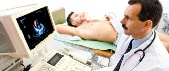
1 ECHO-KG in "MedicCity"
2 Echocardiography at MedicCity
3 Ultrasound of the heart at MedicCity
The essence of echocardiography
Ultrasound of the heart is the process of studying all the main parameters and structures of this organ using ultrasound.
When exposed to electrical energy, the echocardiograph transducer emits high-frequency sound that travels through the structures of the heart, is reflected from them, captured by the same transducer and transmitted to the computer. He, in turn, analyzes the received data and displays it on the monitor in the form of a two- or three-dimensional image.
In recent years, echocardiography has been increasingly used for preventive purposes, which makes it possible to identify cardiac abnormalities at an early stage.
What does an ultrasound of the heart show:
- heart size;
- integrity, structure and thickness of its walls;
- sizes of the cavities of the atria and ventricles;
- contractility of the heart muscle;
- operation and structure of valves;
- condition of the pulmonary artery and aorta;
- pulmonary artery pressure level (to diagnose pulmonary hypertension, which can occur with pulmonary embolism, for example, when blood clots from the veins of the legs enter the pulmonary artery);
- direction and speed of cardiac blood flow;
- condition of the outer shell, pericardium.
1 ECHO-KG in "MedicCity"
2 Echocardiography at MedicCity
3 Ultrasound of the heart at MedicCity
What abbreviations are used in the protocol?
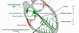
Having received the EchoCG protocol completed by a specialist, the patient is faced with abbreviations that are incomprehensible to him. For example, MPAP is the average pressure in the pulmonary artery, CO and DO are the short and long axis. The most commonly used abbreviations can be seen in the figure.
Basic abbreviations that are indicated in the ultrasound protocol
In most cases, it is impossible to make a diagnosis based only on the results of the protocol. The specialist takes into account such features as ultrasound indicators, the patient’s medical history, the chronology and intensity of the development of symptoms, and other nuances. Taken together, these data help to accurately determine a particular pathology.
Who is prescribed echocardiography?
The following are routinely examined:
- infants - if congenital defects are suspected;
- teenagers are a time of intensive growth;
- pregnant women with existing chronic diseases - to decide on the method of childbirth;
- professional athletes - to monitor the state of the cardiovascular system.
An echocardiogram is required if:
- anomalies of the endocardium and valve apparatus:
- heart tumors;
- arrhythmias;
- threat of a heart attack or a heart attack;
- IHD;
- failures of cardiac activity due to various intoxications;
- attacks of angina pectoris;
- pericarditis of various origins;
- hypertension;
- heart failure.
And also in the process of treating heart diseases, before and after cardiac surgery.
Indications for use
Echocardiography (ultrasound of the heart) is used to diagnose various conditions in a patient. The following symptoms in a person may serve as a reason for the procedure:
- heart murmurs, rhythm disturbances;
- signs indicating the development of heart failure, for example, swelling of the extremities, pain in the liver;
- acute or chronic course of myocardial infarction;
- chronic fatigue, shortness of breath, cyanosis of the skin;
- frequent colds or fever without signs of acute respiratory viral infection;
- predisposition to cardiovascular diseases;
- fainting, angina attacks.
In addition, indications include previous rheumatism, frequent surges in blood pressure, conditions accompanied by pain and numbness in the left arm, shoulder blade, and forearm. The method is used to monitor the effectiveness of treatment for various heart pathologies before upcoming surgery. For the purpose of prevention, it is recommended to have an ultrasound scan for people whose work activities involve frequent emotional or physical stress.
When is a cardiac ultrasound performed?
Indications for cardiac ultrasound are:
- alarming changes in health (increased or interrupted heartbeat, shortness of breath, swelling, weakness, prolonged fever, chest pain, cases of loss of consciousness);
- changes detected in the last ECG;
- increased blood pressure;
- heart murmurs;
- cardiomyopathy;
- manifestations of coronary heart disease;
- heart defects (congenital, acquired);
- pericardial diseases;
- lung diseases.
How to prepare
For classic cardiac ultrasound, no preparation is usually required. But if the patient has high blood pressure (above 160 mmHg) or pulse above 90 beats per minute, it may be necessary to take some medications to avoid errors in interpreting the results. Transesophageal ultrasound is performed strictly on an empty stomach. For intravascular ultrasound, the preparation is the same as for endovascular interventions. In this case, the patient undergoes a special examination, including laboratory tests, an ECG and a consultation with a therapist.
The Euroonko clinic network performs all types of cardiac ultrasound.
We have high-quality equipment that allows us to conduct examinations at an expert level, including endovascular and transesophageal ultrasound, as well as three-dimensional and four-dimensional scanning. Reception is by appointment. Book a consultation around the clock +7+7+78
Preparation for cardiac echocardiography
Ultrasound of the heart does not require special preparations. On the eve of the procedure, the patient is free to eat as usual and perform normal activities. The only thing that will be asked of him is to give up alcohol, caffeine-containing drinks, and strong tea.
If the patient is constantly taking medications, this must be warned in advance so that the results of the study are not distorted.
For each subsequent ultrasound of the heart, you should take a transcript of the previous one. This will help the doctor see the process over time and draw the right conclusions about your condition.
The examination itself takes from 15 to 30 minutes.
The patient, undressed to the waist, is lying on his back or side. A special gel is applied to his chest, ensuring easier sliding of the sensor over the area under study (the patient does not experience discomfort).
The specialist performing echocardiography has access to any areas of the heart muscle - this is achieved by changing the angle of the sensor.
Sometimes standard ultrasound of the heart does not provide complete information about the functioning of the heart, so other types of echocardiography are used. For example, fat on the chest of an obese person can interfere with the passage of ultrasound waves. In this case, transesophageal echocardiography is indicated. As the name suggests, the ultrasound probe is inserted directly into the esophagus, as close to the left atrium as possible.
And to screen cardiac function under stress, the patient may be prescribed stress echocardiography. This study differs from the usual one in that it is performed with a load on the heart, achieved by physical exercise, special medications or under the influence of electrical impulses. It is used primarily to identify myocardial ischemia and the risk of complications of coronary artery disease, as well as for certain heart defects to confirm the need for surgery.
What is electrocardiography and the features of its implementation?
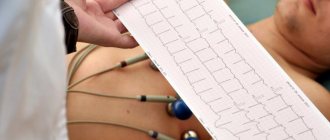
The principle of this examination method is based on recording electronic pulses of the heart that occur during myocardial contraction. The pulses that have arisen are read by the cardiograph, displaying them in the form of a graphic curve on special paper.
Order of conduct:
- Before the procedure, all jewelry should be removed;
- Expose the chest, ankles and hands;
- Lie horizontally and relax;
- Before applying the electrodes, the nurse should treat exposed skin with a cotton pad, which is moistened with water for better conductivity of the impulses.
- Afterwards, 4 electrodes of different colors are applied to the limbs in a certain sequence;
- They are fixed on the chest using suction cups. The number of the latter should be 6 pieces;
- The electrodes are connected to the device, then they are registered.
A cardiogram is necessary when detecting any heart disease; it is also prescribed during a preventive examination.
Using this examination, you can identify such disturbances in the functioning of the heart as:
- Heart rhythm disturbances, for example, tachycardia, arrhythmia, extrasystole;
- Impulse conduction disorder, for example, antiventricular block;
- Myocardial nutritional disorders, for example, heart attack, ischemia;
- Congenital or acquired diseases, for example, disorders in the structure of the fibrous ring, valves, chord;
- Myocardial thickening, for example, atrial ventricular hypertrophy.
Persons who have reached the age of 40 years old must undergo an ECG every year. This will help to identify violations in time at the initial stage.
This study is quite informative, however, despite this, to clarify the diagnosis it is necessary to perform an ultrasound of the heart.
The study is very effective and indicative, but despite this, to clarify the condition of the myocardium, it is necessary to resort to ultrasound diagnostics of the heart.
Pros of the procedure
Among the undeniable advantages are the painlessness of the procedure, safety, and the ability to obtain reliable data. In addition, another advantage is that the procedure does not take much time.
Also, the procedure has no contraindications, and due to the mobility of the device, diagnostics can be carried out outside the clinic.
Disadvantages of the procedure:
The disadvantages of the procedure include the following:
- short recording;
- inability to record heart murmurs;
- impossibility of diagnosing heart defects and various tumors;
- To obtain reliable data, the procedure should be carried out at rest or under load.
Decoding EchoCG
After the examination, the doctor draws up a conclusion. First, a visual picture with the presumed diagnosis is described. The second part of the study protocol indicates the patient’s individual indicators and their compliance with standards.
Decoding the data obtained is not a final diagnosis, since the study can be done not by a cardiologist, but by an ultrasound diagnostic specialist.
It is the cardiologist, based on the collected medical history, examination results, interpretation of tests and data from all prescribed studies, who can draw accurate conclusions about your condition and prescribe the necessary treatment!
Are there any contraindications?
Cardiac echocardiography has no absolute prohibitions, but there are some recommendations that should be followed during diagnosis.
Contraindications:
- An echocardiogram should be performed 2-3 hours after eating. When the stomach is full, the diaphragm may put pressure on the heart, which will affect the accuracy of the data obtained;
- It is recommended to reschedule the procedure for those patients who have open wounds or serious skin diseases in the chest area;
- When the chest is deformed, diagnostic results may be inaccurate.
If transesophageal (TE) echocardiography is performed, it should not be used among patients with an increased gag reflex, mental disorders, or pathologies of the esophagus.
Important! EchoCG is a safe and painless technique, well tolerated by children and adults.
EchoCG norm: what some parameters indicate
There is a range of normal values for one or another cardiac ultrasound indicator for adults (in children the norms are different and directly depend on age).
Thus, along with other important parameters, echocardiography helps to obtain information about the ejection fraction of the heart - this indicator determines the efficiency of the work performed by the heart with each beat.
Ejection fraction (EF) is the percentage of blood volume ejected into the vessels from the heart ventricle during each contraction. If there were 100 ml of blood in the ventricle, and after the heart contracted, 55 ml entered the aorta, it is considered that the ejection fraction was 55%.
When the term ejection fraction is heard, we are usually talking about the left ventricular (LV) ejection fraction, since it is the left ventricle that ejects blood into the systemic circulation.
A healthy heart, even at rest, pumps more than half of the blood from the left ventricle into the vessels with each beat. As the ejection fraction decreases, heart failure develops.
The normal left ventricular ejection fraction for an adult is 55-70%. A value of 40-55% indicates that EF is below normal. An ejection fraction of less than 40% and even lower ejection fractions indicate heart failure in the patient.
What does the specialist see?
During an echocardiogram, the doctor can evaluate the functioning of the heart using several criteria. Each of them has certain norms, and a deviation in one direction or another indicates the presence of various pathologies.
Ultrasound allows you to evaluate the following indicators:
- main characteristics of the heart chambers;
- characteristics of the ventricles and atria;
- functioning of valves and their condition;
- condition of the walls of blood vessels;
- direction and intensity of blood flow;
- characteristics of the heart muscles during the period of relaxation and contraction;
- is there exudate in the pericardial sac?
To make a diagnosis, doctors use certain standards of echocardiography, but sometimes minor deviations in one direction or another are allowed. It depends on the age, weight of the patient and other individual characteristics.
Important! The interpretation of the results obtained should be carried out exclusively by a cardiologist. Once you have the conclusion in your hands, you should not try to establish a diagnosis yourself.
Is cardiac echocardiography safe?
During this study, there is no radiation or other load on the organ. Therefore, if necessary, it can be prescribed even several times a week.
This study is characterized by the absence of complications and side effects.
EchoCG does not harm either the expectant mother or the fetus during pregnancy.
Limitations for the procedure may include inflammatory diseases of the skin of the chest, chest deformities and some other reasons.
Use among pregnant women
During pregnancy, women are susceptible to many diseases. Due to changes occurring in the body, the load on the heart also increases.
Echocardiography is often performed on pregnant women
Indications for echocardiography:
- diabetes;
- hereditary predisposition to heart disease;
- if the patient fell ill with rubella while carrying a baby, or a high concentration of bodies for this disease was detected in the plasma;
- if in the first trimester the woman took any strong medications;
- in the presence of miscarriages in the medical history.
Ultrasounds are often performed on an unborn baby in the womb. The procedure is performed to detect heart defects in the fetus at an early stage and is performed at 18–22 weeks.
How to do an ultrasound of the heart at the MedicCity clinic?
It is advisable for every person to have an echocardiogram at least once in their life. The fairly low cost of cardiac ultrasound is another reason to prefer this particular diagnostic method.
At MedicCity you can undergo all types of cardiac diagnostics - ECG, EchoCG, bicycle ergometry, HOLTER, ABPM, etc.
Just type in a search engine: “Ultrasound of the heart, Savelovskaya metro station, Dynamo metro station.” Or call us at: +7 (495) 604-12-12.
The doctors of our multidisciplinary clinic will do everything possible to relieve your heart pain!
Basic concepts and norms of ultrasound for adults
The heart consists of several sections, each of which plays an important role. Malfunction of any of the chambers can provoke heart failure and other serious complications. The organ consists of the left and right atria, ventricles and valves.
The echocardiographic diagnostic method allows you to visualize the condition of this organ, see the functioning of the valves, the thickness of the myocardium, the speed and direction of blood flow, the presence of vasoconstriction and blood clots in them.
There are no clear boundaries in this area, since each organism is individual. But certain standards still exist. For an adult, the indicators should be as follows:
- in the systole and diastole phase, the wall thickness of the left ventricle is 10–16 and 8–11 mm;
- the wall of the right ventricle should not be dilated and extend beyond the boundaries of 3 to 5 mm;
- interventricular septum in diastole and systole – 6–11 and 10–15 mm;
- aortic circumference – from 18 to 35 mm;
- in women and men, the total myocardial mass should be between 90–140 g and 130–180 g;
- heart rate – 75–90;
- the ejection fraction should not be less than 50%.
In addition, such parameters are assessed in adult patients as the volume of fluid in the heart sac (35 sq. ml), the diameter of the aortic valve should not exceed one and a half centimeters, and the opening of the mitral valve (4 sq. cm).
