First aid for hypovolemic shock
Hypovolemic shock is a pathological condition that occurs as a result of a rapid decrease in the volume of blood circulating in the body.
Causes of hypovolemic shock:
- acute blood loss;
- burn disease;
- dehydration (uncontrollable vomiting, profuse diarrhea);
- gestosis;
- polyuria;
- endocrine disorders;
- uncontrolled use of certain medications.
In the development of hypovolemic shock, three phases are distinguished: BCC deficiency (circulating blood volume), stimulation of the sympathoadrenal system, shock.
Classification of hemorrhagic shock
The classification of hemorrhagic shock is based on the stage of development of the pathological process, according to which there are 4 degrees of hemorrhagic shock:
- First degree shock (compensated reversible shock). It is caused by a small amount of blood loss, which is quickly compensated by functional changes in the work of cardiovascular activity.
- Shock of the second degree (subcompensated). Developing pathological changes are not fully compensated.
- Third degree shock (decompensated reversible shock). Disturbances are expressed in various organs and systems.
- Fourth degree shock (irreversible shock). It is characterized by extreme depression of vital functions and the development of irreversible multiple organ failure.
Causes of hypovolemic shock
Burns. Hypovolemia is promoted by the loss of plasma during extensive burns, aggravated by intoxication with tissue decay products.
Kidney diseases. The causes of hypovolemic shock can be excessive excretion of water and sodium, as well as the presence of glucose in the urine, which carries a significant amount of fluid.
Excretion of water by the intestines. We are talking about infections accompanied by profuse diarrhea. Emergency care for hypovolemic shock will be discussed further.
Blood loss is the most common cause when blood leaks into the lumen of a tissue (organ) or into the external environment.
Degrees of hypovolemic shock:
- first (mild) – loss of up to 15% of bcc (blood pressure and breathing are normal, possible pallor of the skin, anxiety);
- second (moderate) - loss of blood volume from 30 to 40% (blood pressure drops below 100 mm Hg, rapid pulse, pale skin, increased sweating, severe anxiety);
- Third (severe) – blood loss of more than 40% (weak and rapid pulse, blood pressure below 70 mm Hg, pale, sweaty skin, incoherent speech, loss of consciousness).
Detailed information about the treatment of hypovolemic shock is available on the pages of our website Dobrobut.com. Use the special form and make an appointment with the right specialist. At the clinic you can undergo a full examination of the body and a course of rehabilitation after serious illnesses.
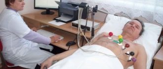
Diagnostics
In diagnosis, it is important to determine the type of fluid loss. If there is or information about bleeding, vomiting, diarrhea, or a large burn surface, the symptoms themselves indicate the root cause of the pathological disorders. The doctor experiences significant difficulties if the bleeding is internal with an unclear cause.
The patient should be taken to the hospital as quickly as possible. Here you must take:
- blood tests;
- the group and Rh factor are determined;
- OCC;
- urine is examined for specific gravity (an indicator of concentration), protein and red blood cells.
X-rays are taken to identify hidden fractures.
If blood in the abdominal cavity is suspected, laparoscopy is necessary.
During treatment, the electrolyte composition and alkaline balance are examined. These indicators are important for choosing solutions of the desired concentration and composition.
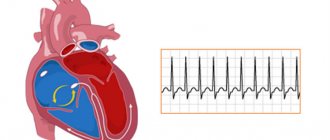
Hemorrhagic shock is considered a type of hypovolemic shock. It is practically important to determine the amount of blood loss. There are different ways to do this.
Calculation of the shock index by dividing the pulse rate by the upper pressure: if normally this coefficient is about 0.54, then with shock it increases.
To establish blood loss during fractures in an adult, average values are used depending on the type:
- femur fracture - 1 l;
- shin bones - about 750 ml;
- shoulder - up to 500 ml;
- pelvic bones - up to 3 liters.
When examining the chest organs, radiologists approximately determine the amount of blood that has spilled into the pleural cavities:
- if you can clearly see the liquid level - up to 0.5 l;
- when the fields of lung tissue are darkened - up to 2l.
When examining a patient with suspected internal bleeding into the abdominal cavity, the surgeon focuses on the symptom of fluid leakage. This means that there is at least a liter of liquid in the cavity.
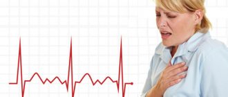
Signs of hypovolemic shock
Obvious symptoms of this condition develop with a loss of 10-20% of the blood volume. Early signs of hypovolemic shock include:
- thirst;
- anxiety, irritability;
- pale skin covered with sticky sweat;
- decreased blood pressure, rapid breathing;
- dizziness;
- confusion (not always).
In the absence of adequate medical care, the patient's condition worsens and late symptoms develop: a sharp decrease in blood pressure, severe tachycardia, decreased temperature, dizziness, loss of consciousness.
Treatment of hypovolemic shock is usually carried out by a resuscitator in collaboration with specialized specialists. Therapy is aimed at eliminating respiratory and circulatory hypoxia and ensuring sufficient blood supply to organs, as well as restoring circulating blood volume.
Meningococcal infection
Meningococcemia is one of the forms of generalized meningococcal infection, characterized by an acute onset with a rise in body temperature to febrile levels, general intoxication, hemorrhagic skin rashes, and the development of infectious-toxic shock.
Clinical manifestations of meningococcal infection:
- Often acute onset or sharp deterioration due to nasopharyngitis.
- High, persistent fever with signs of impaired peripheral circulation. Early signs of fulminant toxic progression of meningococcal infection may be a “two-humped” nature of the temperature curve - the first increase in body temperature to 38.5ºC is reduced with the help of antipyretics, the second (after 9-18 hours) - to 39.5-40ºC without the effect of antipyretics.
- Hemorrhagic rash that appears a few hours or 1-2 days after the onset of fever. The typical localization of the rash is the outer surface of the thighs and legs, buttocks, feet, hands, and lower abdomen. Often a hemorrhagic rash is preceded or combined with a polymorphic roseolous or roseolous papular rash with the same localization, less often on the face.
- General cerebral symptoms: intense headache, “cerebral vomiting”, possible disturbances of consciousness, delirium, hallucinations, psychomotor agitation, convulsions.
- Meningeal syndrome. It usually appears later, against the background of advanced symptoms of meningococcemia.
Threatening syndromes in generalized forms of meningococcal infection are:
- infectious-toxic shock develops during the hyperacute course of meningococcemia. As a rule, symptoms of shock occur simultaneously or after the appearance of a hemorrhagic rash. However, ITS can occur without a rash, so all children with infectious toxicosis should have their blood pressure measured;
- cerebral edema with brainstem dislocation. It manifests itself as impaired consciousness, hyperthermia, severe meningeal symptoms (sometimes their absence in the terminal stage of the disease), convulsive syndrome and unusual changes in hemodynamics in the form of relative bradycardia and a tendency to increase blood pressure. In the terminal stage of cerebral edema there is absolute bradycardia and respiratory arrhythmia.
Urgent Care:
- Ensure patency of the upper respiratory tract, oxygen therapy.
- Administer chloramphenicol (chloramphenicol succinate) at a dose of 25-30 mg/kg intramuscularly.
- Intravenous (if not possible, intramuscular) administration of glucocorticoids in terms of prednisolone 5-10 mg/kg or dexamethasone 0.6-0.7 mg/kg body weight; if there is no effect and it is impossible to transport the patient, it is necessary to re-introduce hormones in the same doses (for ITS stage III, the dose of corticosteroids can be increased 2-5 times).
- Introduce a 50% solution of metamizole (analgin) 0.1 ml/year of life intramuscularly + 1% solution of diphenhydramine (diphenhydramine) at a dose of 0.1-0.15 ml per year of life intramuscularly (or 2% solution Suprastin solution – 2 mg/kg intramuscularly) + 2% papaverine solution 0.1 ml/year of life).
- For agitation, convulsive syndrome - 0.5% diazepam solution in a single dose of 0.1 ml/kg body weight (no more than 2 ml per administration) intravenously or intramuscularly, for intractable convulsions again in the same dose .
- In case of severe meningeal syndrome - intramuscular administration of a 1% furosemide solution at the rate of 1-2 mg/kg intramuscularly or 25% magnesium sulfate solution 1.0 ml/year of life intramuscularly.
Call the resuscitation team for yourself!!! Hospitalization in the intensive care unit of the nearest hospital. If meningococcal infection is suspected, hospitalization is indicated .
Intestinal toxicosis with exicosis is a pathological condition that is a complication of acute intestinal infections (AI) due to exposure to toxic products and significant fluid losses on the body, which leads to disruption of hemodynamics, water-electrolyte metabolism, acid-base reserve, and the development of secondary endogenous intoxication.
In the pathogenesis of toxicosis with exicosis, the leading role is played by dehydration, which leads to a deficiency in the volume of extra- (and in severe cases, intra-) cellular fluid and the volume of circulating blood. The degree of exicosis (dehydration) determines the choice of therapeutic tactics and affects the prognosis.
Severity of dehydration
| Signs | Degrees of dehydration | |||
| I degree | II degree* | III degree | ||
| Underweight | Up to 5% | 6-9 % | More than 10% | |
| BCC deficiency | More than 10% | 11-20 % | More than 20% | |
| General state | good | Excitation | Lethargic, lethargic, unconscious | |
| Chair | Infrequent (4-6 times a day) | Up to 10 times a day | Frequent (more than 10 times a day), watery | |
| Vomit | One-time | Repeated (3-4 times a day) | Multiple | |
| Thirst | Moderate | Sharply expressed | Refusal to drink | |
| Skin fold | Spreads out quickly | Unfolds slowly (≤2 sec) | Unfolds very slowly (>2 sec) | |
| Mucous membranes | Moist or slightly dry | Dry | Very dry, bright | |
| Cyanosis | ||||
| Eyeballs | Without features | Soft | They're sinking | |
| Voice | Without features | Weakened | Aphonia | |
| Heart sounds | Loud | Slightly muted | Deaf | |
| Heart rate | Norm | Moderate tachycardia | Severe tachycardia | |
| Diuresis | Saved | Reduced | Significantly reduced | |
| Blood plasma electrolytes | Norm | Hypokalemia | Hypokalemia | |
| CBS | Norm | Compensated acidosis | Decompensated acidosis | |
| Rehydration method | Oral | Parenteral | ||
- II degree of exicosis is divided into IIA and IIB (with loss of body weight 6-7% and 8-9%, respectively).
Measures for dehydration
When starting to rehydrate children with exicosis, it is necessary to determine which route (orally and/or parenterally) and what volume of fluid should be administered to the child, at what speed to administer, and what solutions to use. Oral rehydration is carried out for exicosis of I, IIA degrees, parenteral - for exicosis of IIB and III degrees.
Outpatient treatment is possible (in the absence of other contraindications, see below) for grade I exicosis with oral rehydration.
Oral rehydration therapy
Indications: exicosis I, IIA degrees.
- The necessary solutions are glucose-salt (rehydron, oralit, gastrolit, glucosalan). In children under 3 years of age, it is advisable to combine glucose-saline solutions with salt-free solutions (tea, water, rice water, etc.) in a ratio of 1:1 - for severe watery diarrhea, 2:1 - for loss of fluid mainly through vomiting, 1: 2 – with loss of fluid with perspiration. The administration of saline and salt-free solutions alternates.
The amount of liquid for each hour of administration is divided into ½ teaspoon - 1 tablespoon (depending on age) every 5-10 minutes. If there is vomiting, rehydration does not stop, but is interrupted for 5-10 minutes and then continues again.
Features of the introduction of solutions at the 1st stage (the first 6 hours from the start of treatment):
- for grade I dehydration: 50 ml/kg for 6 hours. + 10 ml/kg b.w. after each loose stool or vomiting;
- for degree IIA dehydration: 80 ml/kg for 6 hours + 10 ml/kg b.w. after each loose stool or vomiting.
At the 2nd stage, maintenance therapy is carried out in the amount of ongoing fluid losses administered at this stage (80-100 ml/kg per day until the loss stops).
Therapy for hypovolemic shock
Hypovolemia requires immediate treatment. The patient is usually hospitalized in the intensive care unit, where complex therapy will be carried out aimed at restoring circulating blood volume, normalizing blood circulation in vital organs (brain, heart, lungs, kidneys) and restoring the acid-base balance. For this purpose, infusion therapy, blood replacement drugs, glucocorticosteroids, and sedatives are prescribed. In parallel, oxygen inhalations are carried out, and if necessary, the patient is transferred to mechanical ventilation.
Emergency care for hypovolemic shock before the ambulance arrives:
- In an unconscious state, a person must be placed on his back and his head turned to the side (in the absence of damage to the cervical spine);
- raise your legs 30 cm;
- cover with a blanket (to avoid hypothermia).
Clinical recommendations for providing first aid for hypovolemic shock in children are identical to those described above.
The importance of fluid in human physiology
Water is part of the entire complex of fluids that wash organs and tissues. It is the main component of blood, lymph, cerebrospinal and interstitial fluid, secretions of the salivary glands, gastric and other juices produced by internal organs, tears, and urine.
Liquid creates a universal internal environment for the existence of cells. Through it the following is carried out:
- nutrition and removal of toxins;
- “orders” are delivered from the nervous and endocrine centers;
- the necessary brain structures are excited.
The role of liquid in ensuring metabolic processes is great. All biochemical reactions take place only in dissolved form. By changing the amount of water in the body in accordance with physiological needs, the required concentration of salts, electrolytes, acids and alkalis, biological substances, everything that ensures the internal environment or homeostasis is maintained.
The preservation of homeostasis indicators is guaranteed by natural tissue barriers (skin, mucous membranes of organs and blood vessels). Equilibrium can change under the influence of regulatory systems, but within very narrow limits.
Therefore, any disturbances in the composition of liquid media can be used to judge the pathology that has arisen. A decrease in fluid causes significant changes in homeostasis: some substances are lost along with water, others sharply increase in concentration. Pathophysiological disorders may concern:
- cellular composition of blood;
- alkaline balance;
- concentration of dissolved substances.
The functional capacity of systems depends on the norms of water distribution in the body.
Changed conditions cause many diseases.
In humans, it is convenient to judge the volume of fluid by the indicator of circulating blood. It is calculated in the laboratory. A decrease of 25% in healthy people is well compensated and does not cause any significant changes in homeostasis. 90% of the blood is in the vascular bed, the rest is deposited in the spleen and bones. If necessary, it is thrown out of storage and replenishes losses.
Large losses lead to varying degrees of hypovolemia, and in the absence of compensation and assistance - to a hypovolemic shock state.
Symptoms
The clinical picture of hemorrhagic shock develops in accordance with its stages. Clinically, signs of blood loss come to the fore. At the stage of compensated hemorrhagic shock, consciousness, as a rule, does not suffer, the patient notes weakness, may be somewhat agitated or calm, the skin is pale, and the extremities are cold to the touch.
The most important symptom at this stage is the desolation of the subcutaneous venous vessels in the arms, which decrease in volume and become thread-like. The pulse is weak and rapid. Blood pressure is usually normal, sometimes elevated. Peripheral compensatory vasoconstriction is caused by overproduction of catecholamines and occurs almost immediately after blood loss. Against this background, at the same time the patient develops oliguria . At the same time, the amount of urine excreted may be reduced by half or even more. Central venous pressure decreases sharply, which is due to a decrease in venous return. In compensated shock, acidosis is often absent or local in nature and mildly expressed.
At the stage of reversible decompensated shock, the signs of circulatory disorders continue to deepen. In the clinical picture, which is characterized by signs of the compensated stage of shock (hypovolemia, pallor, profuse cold and sticky sweat, tachycardia, oliguria), the main cardinal symptom is hypotension , which indicates a disorder in the circulatory compensation mechanism. It is in the stage of decompensation that organ circulatory disorders (in the intestines, liver, kidneys, heart, brain) begin. Oliguria, which in the compensation stage develops as a result of compensatory functions, at this stage occurs on the basis of a decrease in hydrostatic blood pressure and renal blood flow disorders.
At this stage, the classic clinical picture of shock appears: acrocyanosis and coldness of the extremities, increased tachycardia and the appearance breath , dullness of heart sounds, which indicates a deterioration in myocardial contractility. In a number of cases, there is a loss of individual or a whole group of pulse impulses in the peripheral arteries and the disappearance of heart sounds during deep inspiration, which indicates an extremely low venous return.
The patient is inhibited or in a state of prostration. Shortness of breath and anuria develop . DIC syndrome is diagnosed. Against the background of the most pronounced vasoconstriction of peripheral vessels, direct discharge of arterial blood into the venous system occurs through opening arteriovenous shunts, which allows increasing oxygen saturation of the venous blood. At this stage, acidosis is pronounced, which is a consequence of increasing tissue hypoxia .
The stage of irreversible shock is not qualitatively different from decompensated shock, but is a stage of even more pronounced and profound disorders. The development of a state of irreversibility is manifested by a matter of time and is determined by the accumulation of toxic substances, the death of cellular structures, and the appearance of signs of multiple organ failure. As a rule, at this stage there is no consciousness, the pulse in the peripheral vessels is practically not determined, blood pressure (systolic) is at the level of 60 mm Hg. Art. and lower, difficult to determine, heart rate at 140/min., breathing is weakened, rhythm is disturbed, anuria. There is no effect from infusion-transfusion therapy. The duration of this stage is 12-15 hours and ends in death.
During pregnancy
Hemorrhagic shock in obstetrics, which occurs during pregnancy, during labor, as well as in the early/late afterbirth period, is one of the significant causes in the structure of maternal mortality, accounting for about 20–25%. The rate of obstetric hemorrhage relative to the total number of births varies between 5-8%.
The specifics of bleeding in obstetrics are:
- The suddenness of their appearance and massiveness.
- High risk of fetal death, which necessitates urgent delivery until stable stabilization of hemodynamic parameters and completion of infusion-transfusion therapy in full.
- Combination with pronounced pain syndrome.
- Rapid depletion of compensatory and protective mechanisms. At the same time, the risk is especially high in pregnant women with late gestosis and in women with complicated labor.
The permissible blood loss during childbirth during its normal course should not exceed 250-300 ml (approximately 0.5% of a woman’s body weight). This amount of blood loss refers to the “physiological norm” and does not negatively affect the condition of the woman in labor. The main causes of acute pathological blood loss during pregnancy with the development of hemorrhagic shock are: ectopic pregnancy , placental abruption/previa, multiple pregnancy, complications during childbirth, cesarean section, uterine rupture.
Features of hemorrhagic shock in obstetric pathology include: its frequent development against the background of a severe form of gestosis in pregnant women, the rapid development of hypovolemia , disseminated intravascular coagulation syndrome, arterial hypotension , and hypochromic anemia .
In case of HS, which developed in the early postpartum period against the background of hypotonic bleeding, a short period of unstable compensation is characteristic, after which irreversible changes quickly develop (persistent hemodynamic disturbances, DIC syndrome with profuse bleeding, respiratory failure and impaired blood coagulation factors with activation of fibrinolysis).