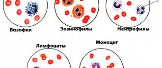
Despite the frequent use of the term “hypovolemia,” many doctors do not have an accurate idea of its meaning. Obviously, this is why they consider it possible not to use objective criteria when voicing this diagnosis; as a result, it turns into a subjective opinion. Moreover, slang expressions such as “underfilled” or “overfilled” are used in relation to the patient, referring to defects in infusion therapy.
Meanwhile, a violation of the patient’s volemic status is always a very serious symptom of trouble in the body, requiring objective confirmation and immediate therapeutic response, since volemia is the foundation of circulation, and therefore of exchange, that is, the very existence of the body. That is why a free interpretation of volemia can and does lead to great losses for the patient.
The terms “hypovolemia” and “hypervolemia” imply the presence of normovolemia, in comparison with which a decrease or increase in blood volume is determined. The search for the very concept of “normovolemia” continued for more than a hundred years, and now, finally, an almost generally accepted decision has appeared to consider normal only that volume of circulating blood (CBV), which in size corresponds to the capacity of the vascular bed. In this case, relaxed hemodynamics and breathing are observed, that is, there is no pronounced tachycardia, peripheral vasospasm, or increased respiratory movements.
Hypovolemia: a dangerous condition that requires immediate attention
What do we know about hypovolemia?
Hypovolemia is a condition when, as a result of fluid loss, the volume of blood that circulates in the human body decreases.
This violation poses a serious threat to the health and life of the patient. A severe degree can lead to hypovolemic shock and the development of multiple organ failure. Therefore, as a rule, the patient requires immediate medical attention: hospitalization and intensive care. Treatment is carried out in a hospital setting.
In rare cases, hypovolemia is caused not by the loss of fluid, but by its redistribution in the tissues of the body. At the same time, the filling of blood vessels with blood is reduced. This phenomenon is called relative hypovolemia. Blood loss and dehydration are absolute.
Reasons and mechanism of formation
The pathological hypovolemic process is based on severe dehydration.
Leads to it:
- Vomiting, characteristic of withdrawal syndrome after binge drinking.
- Diarrhea, with the help of which the body cleanses itself of alcohol toxins.
- Liver diseases characteristic of alcoholism (toxic hepatitis and cirrhosis). In this case, non-functional leakage of plasma into the intercellular space occurs due to a decrease in the oncotic pressure of the blood.
- Insufficient intake of fluid and protein into the body.
Hypovolemia is preceded by increased venous and hydrostatic pressure in small arteries, reflecting the negative influence of factors predisposing to a painful condition. Secondarily, the permeability of the vascular wall increases. Sweating of blood through arteries and veins leads to its general deficiency.
The body tries to compensate by:
- Producing the required amount of plasma.
- Slowing venous return.
- Spasms of small vessels.
The protective reaction allows you to maintain the activity of the brain and heart in a relatively normal state for some time and avoid circulatory complications.
What causes the condition
Most cases are associated with severe dehydration, blood loss, and large burns.
Dehydration can be caused by:
- repeated vomiting and diarrhea, excessive sweating
- insufficient fluid intake,
- taking medications, such as diuretics.
Hypovolemia that is caused by loss of plasma rather than blood is called polycythaemic.
Blood loss occurs as a result of injury, surgery, or internal bleeding. Blood loss associated with internal bleeding is considered especially dangerous, since it is not always possible to immediately determine where it is localized.
Massive blood loss is caused by:
- gastrointestinal bleeding,
- rupture of the fallopian tube during ectopic pregnancy,
- rupture of an aortic aneurysm.
A general decrease in blood volume is called normocythemic hypovolemia.
4.Treatment
Depending on the situation, urgent resuscitation measures are taken. The primary objectives are to stop bleeding, restore breathing and replace blood loss through blood transfusion or transfusion of donor blood, and, if necessary, to relieve pain shock. In the future, measures are needed to normalize the acid-base balance, prevent thrombosis (DIC syndrome), prevent renal failure and secondary infections. Further management of the patient is determined by the results of the diagnostic examination and the dynamics of the condition.
Symptoms
The condition can be recognized by the following signs:
- dry mucous membranes
- pale skin
- decreased skin turgor
- rapid breathing
- lowering blood pressure levels
- tachycardia
- anxiety, confusion
- weak pulse (due to decreased cardiac output)
- thirst
- dizziness
- lack of urination for a long time.
Acute hypovolemia leads to an emergency condition - hypovolemic shock, which can result in coma.
Content:
- Causes and mechanism of development
- Classification
- Signs of hypovolemic disorder
- Treatment of hypovolemia
Hypovolemia is a decrease in blood volume with the development of alcohol withdrawal syndrome. This process occurs due to the recombination of fluid, due to its transition from the venous bed into the cells and the space between them. This phenomenon occurs during the accumulation of toxic substances inside tissues due to dysfunction of the enzyme system, coronary artery disease, and poor functioning of organs. In addition to perfusion problems, a decrease in circulating blood volume (CBV) is manifested by an inadequate ratio of systolic and diastolic blood pressure and a drop in cardiac output. Developing acute pathology is life-threatening and requires immediate medical attention.
Diagnostics
The main means of establishing a diagnosis is examination and interview (if possible) of the patient.
If hypovolemia is caused by visible trauma, large-area burns, its diagnosis is not difficult.
If a patient is suspected of dehydration or internal bleeding, the patient is sent for a clinical and biochemical blood test, a urine test, which will determine whether there is a change in the water-electrolyte balance.
Also, to clarify the diagnosis, the doctor performs CT, X-ray, ultrasound and endoscopy of organs in which bleeding may develop.
Pathogenesis
Questions immediately arise: what does the capacity of the vascular bed correspond to, how does it change, can it be measured and compared with something to determine its adequacy? How is the correspondence of the BCC to the capacity of the vascular bed expressed?
There is a clear answer to the first question: the capacity of the vascular bed normally corresponds to the level of metabolism in the body. This capacity often changes in accordance with changes in the level of exchange (the BCC also often changes). Obviously, it can be compared with the amount of absorbed oxygen per unit time. We do not yet know how to measure the capacity of the vascular bed, but we know how to evaluate its decrease - it is expressed in peripheral vasoconstriction, which determines the degree of such decrease. The same vasoconstriction in peripheral tissues or centralization of blood circulation is a measure of the discrepancy between the BCC and the capacity of the vascular bed. In other words, with a decrease in BCC, a spasm of peripheral vessels occurs, which reduces the capacity of the vascular bed and thereby brings the reduced BCC into full or partial correspondence with the reduced capacity of the vascular bed.
In this way, the most serious circulatory disturbance is prevented: a decrease in venous return and, consequently, cardiac output. It can be considered that the centralization of blood circulation and at least partial restoration of the correspondence between the bcc and the capacity of the vascular bed are the most important compensatory reaction for maintaining functioning central and peripheral hemodynamics in conditions of a decrease in bcc (blood loss, plasma loss, exsiccation). At the same time, we are able to judge by the state of peripheral blood circulation the dynamics of the bcc and its correspondence to the capacity of the vascular bed: a reduction in peripheral blood flow demonstrates hypovolemia, normal peripheral blood flow indicates the correspondence of the bcc and the capacity of the vascular bed.
Now it remains to establish a method for practical assessment of volemia based on the state of peripheral blood flow. This can be done both by the dynamics of the temperature of peripheral tissues and by the rate of urine formation by the kidneys, but the simplest and most reliable way to assess peripheral blood flow is by the dynamics of the amplitude of the plethysmographic curve (reflecting the dynamics of the volume of the organ in connection with the cardiac cycle). The plethysmographic curve is taken during pulse oximetry. The difficulty lies not in recording the photoplethysmographic curve, but in its interpretation. The fact is that increased sympathetic tone and spasm of peripheral vessels occur not only due to hypovolemia, but also with painful stimulation (inadequate analgesia), decreased body temperature, hypocapnia (hyperventilation), and decreased cardiac output. If we exclude the influence of all the listed factors on the amplitude of the pulse (plethysmographic) curve, we can talk about control of volemic status based on the dynamics of the amplitude of this curve. To do this, you need to have data on body temperature, capnometry, symptoms of the adequacy of anesthesia and, finally, an ECG. Experience shows that such an exception is quite possible when using appropriate monitors.
Maintaining normal peripheral blood flow and controlling it is the most important condition for the successful course of surgery, anesthesia, and the postoperative period; resolution of other pathological conditions. Whatever the cause of the disturbance in peripheral blood flow, a diagnosis must be made and therapy must be carried out to eliminate the effect of the disturbing factor. So monitoring the plethysmographic curve is needed not only to assess the patient’s volumetric status, but also to prevent disturbances in the peripheral blood supply associated with other factors.
Hypovolemia, or a discrepancy between the volume of circulating blood and the capacity of the vascular bed, can also occur with normal BCC due to an increase in the capacity of the bed. The described hypovolemia occurs with severe allergic reactions, endogenous and exogenous intoxications. This type of hypovolemia is sometimes called relative; it is accompanied by a decrease in venous return, cardiac output and, consequently, a disturbance in peripheral blood flow and supply to peripheral tissues. The completion of the process is hypoxia.
Thus, different types of hypovolemia are based on different vascular reactions (peripheral spasm with a decrease in blood volume and vascular relaxation with an increase in the capacity of the vascular bed), different pathogenesis, and different means are used for their treatment (replenishment of blood volume in one case and an increase in vascular tone in another), but in both cases hypoxia develops as an integral expression of shock: in one case, hypovolemic, in the other, vasogenic.
After objective (symptoms of impaired peripheral blood flow) confirmation of hypovolemia with a decrease in blood volume, it is necessary to find out the cause of this decrease and, in accordance with it, urgently begin treatment of the patient.
There are several known main reasons for the development of a decrease in the volume of circulating blood (blood loss, burn disease, release of the liquid part of the blood into the so-called third space and exsiccation - insufficient replenishment of fluid losses. Each of these reasons determines the nature of hypovolemia, its consequences and, most importantly, treatment.
Hypovolemia in children
Danger of condition
The child's body is not sufficiently adapted to compensate for fluid deficiency. This feature should be taken into account when hypovolemia is suspected, since a state of shock in a child can occur much faster than in an adult. This is even more important the younger the child is. Thus, in children up to one year old, the ductus arteriosus may remain open, which, when fluid or blood is lost, actually deprives the lungs of blood supply. Therefore, the issue of hospitalization of the child is resolved immediately.
Causes
In newborns, hypovolemia can develop due to blood loss through the placenta, internal bleeding as a result of injury, or fluid accumulation in the abdominal or pleural cavity.
In older children, in addition to blood loss, the condition is caused by uncontrollable vomiting and diarrhea due to intestinal infections and intoxication; non-compliance with drinking regime.
Treatment
Treatment is prescribed based on the patient's condition.
Mild degrees require fluid replacement (drip, intravenous).
If the cause is injury, the first thing to do is stop the bleeding. Then restore normal hemodynamic parameters and breathing, while simultaneously treating the injury and pain relief for the patient.
Significant blood loss requires transfusion of plasma, red blood cells, platelets, and donor blood. Further therapy is aimed at preventing thrombosis, associated infections, and preventing renal dysfunction.
Signs of hypovolemic disorder
Symptoms of the disorder depend on its severity.
In the practice of narcology, the following stages of hypovolemia are distinguished:
- Easy. Minor fluid losses (up to 20% of bcc) are accompanied by a decrease in blood pressure, increased heart rate 10-15 beats higher than normal, and slight shortness of breath. The patient is pale, complains of weakness, dry mucous membranes, dizziness, and attacks of nausea.
- Average. The patient loses up to 40% of his total blood volume. In this case, the pressure is sharply reduced (below 90 mm Hg) and the pulse is above 120-130 beats per minute. Breathing is sharply rapid and intermittent. Externally, pallor and cyanosis of the skin in the area of the nasolabial triangle are revealed. There is severe sweating, lethargy, and decreased urine output due to severe thirst.
- Heavy. With a large-scale decrease in blood volume (above 40%), a critical drop in blood pressure is detected. The pulse is barely detectable and reaches 150 or more beats per minute. No urine is released. The patient's appearance indicates that he is in critical condition. There is deathly pale skin, a pointed face, sunken cheeks, and an unconscious state.
Loss of BCC is often accompanied by:
- Hypoglycemia is a serious complication of alcohol withdrawal syndrome. A harbinger of the development of hypoglycemia is any kind of liver disease, a decrease in glycogen reserves, and poor non-caloric nutrition.
- Hyponatremia is a disorder most often found in patients who abuse low-alcohol drinks (cocktails, beer). In this condition, peripheral edema occurs in combination with hypovolemia.
- Hypernatremia is a condition that occurs with encephalopathy, acute liver failure, and a clear deficiency in the amount of circulating blood.
- Hypomagnesemia is a deficiency of magnesium inside cells in people with alcoholism, regardless of its content in the plasma. The most common symptoms: general weakness of the body and drowsiness. The risk of seizures and heart problems (palpitations) may increase.
- Hypokalemia. In people suffering from alcoholism, as a rule, potassium deficiency is detected in the body, even if its content in the blood plasma is at normal levels. In combination with an overexcited nervous system, the lack of this element leads to irreparable disturbances in the functioning of the heart with a threat to life. Anyone suffering from alcohol withdrawal syndrome is prescribed potassium supplements.
This pathology manifests itself:
- Fatigue quickly.
- Depression.
- Attacks of tachycardia.
- Muscle weakness.
Alcohol withdrawal syndrome may be accompanied by metabolic acidosis, which occurs due to the accumulation in the body of breakdown products of alcohol and other substances of pathological metabolism, in the process of tissue hypoxia and respiratory alkalosis.
Drugs for hypovolemia
Treatment is carried out in a hospital, often in the intensive care unit. In most cases, infusion therapy is used.
The drugs of choice are solutions of colloids (solutions of gelatin and dextran, for example, rheopolyglucin) and crystalloids (Ringer's solution), blood substitutes (voluven, refortan).
To avoid infections, the patient is prescribed broad-spectrum antibiotics.
To raise the patient's blood pressure, norepinephrine and dobutamine are prescribed.
Clinical manifestations, diagnosis and treatment of hypervolemia
Signs of hypervolemia and treatment tactics largely depend on its type and the reasons that caused this condition.
In the case of physiological and functional reasons that do not go beyond the adaptive abilities of our body, the manifestations are short-term and without any special medical manipulations the body will independently restore its normal state.
If hypervolemia is caused by any chronic or acute disease, treatment tactics are aimed primarily at the disease itself causing an increase in the amount of intravascular blood, and also, if necessary, at relieving the immediate symptoms of hypervolemia, which manifests itself in a variety of ways and nonspecifically:
- Increased blood pressure;
- An increase in the load on the heart can lead to manifestations of heart failure and angina pectoris;
- unexplained weight gain;
- Edema;
- Dyspnea;
- Feeling of dry skin and dry mouth;
- Urinary disorders;
- Increased breathing rate and feeling of heaviness when breathing;
- General weakness;
- Headache;
- Pain in the lumbar region;
- Increased fatigue.
Diagnosis of a hypervolemic state in practical medicine is difficult, which is due to the lack in clinical practice of objective, reliable, and most importantly safe methods for determining the volume of circulating blood. In other words, the methods that are used have proven themselves well in experimental science, explained this pathological process and laid the scientific foundations for the treatment of hypervolemia. Only the hematocrit indicator remains available for practical use, which is of great importance in determining the type of hypervolemia and the reasons that caused it.
Treatment tactics are based on two directions:
Etiotropic (directed at the cause of the pathology) treatment:
- Fighting kidney diseases;
- Surgical treatment of heart defects as early as possible;
- Treatment of endocrine diseases;
- Fighting tumors and congenital diseases of the blood system;
- Treatment of acute and chronic lung diseases;
- Carefully monitor the volume of intravenous infusions.
Symptomatic (aimed at combating the manifestations of pathology) treatment:
- High blood pressure is treated with the use of antihypertensive drugs with an emphasis on diuretics;
- Angina caused by hypervolemia requires, first of all, a reduction in the load on the heart and only then the use of antianginal drugs;
- One of the leading elements of help with hypervolemia is placing the patient in comfortable conditions with normal ambient temperature and a sufficient amount of oxygen in the inhaled air.
Traditional medicine can also be considered effective and gentle treatment methods:
- Hirudotherapy (use of leeches) has a direct effect on blood volume, reducing it, and also reduces blood viscosity and slightly reduces the number of formed elements, which can normalize hematocrit in polycythemic hypervolemia;
- Herbal diuretics: fennel, dill, viburnum, bearberry, horsetail and others.
Treatment and especially diagnosis of hypervolemia requires a careful, comprehensive approach on the part of a qualified doctor, since the apparent simplicity and harmlessness of this condition may hide the initial manifestations of a serious disease, the early and timely diagnosis of which can save a person’s health and even life.
List of used literature
- Savelyev V.S. Comparative effectiveness of plasma substitutes for normovolemic hemodilution and correction of acute blood loss / V.S. Savelyev, N.A. Kuznetsov // Vestn. surgery. –1985.
- Bryusov P.G. Emergency infusion-transfusion therapy for massive blood loss / P.G. Bryusov // Hematology and transfusiology. –1991. – No. 2.
- Pshenisnov K.V., Aleksandrovich Yu.S. Massive blood loss in pediatric practice. Hematology and transfusiology. 2020;65(1):70-86
- Pleskov A.P. Invasive monitoring of central hemodynamics. // Intensive care, ed. V.D.Malysheva, M.: Medicine, 2002. P. 175–190.
Frequently asked questions about hypovolemia
What diseases can cause hypovolemia?
The condition occurs with gastrointestinal diseases, accompanied by uncontrollable vomiting and diarrhea, kidney disease, sharp drops in blood glucose levels, and some allergic reactions. However, hypovolemia is most often caused by blood loss or extensive burns.
How does hypovolemia manifest?
The patient has dry mucous membranes, tachycardia, thirst, and impaired skin turgor. As the condition worsens, a drop in blood pressure, weak pulse, and rapid breathing occur. In the acute stage - confusion.
How is hypovolemia treated?
The main method of treatment is fluid replacement. In most cases, intravenous administration of saline or electrolyte solutions is used.
Operative (controlled) blood loss
Above, we discussed methods for diagnosing a decrease in blood volume based on manifestations of peripheral blood flow disorders. These methods are indispensable in assessing the degree of hypovolemia, which was a consequence of community-acquired blood loss, blood loss to internal organs and tissues (for example, ulcerative bleeding in the stomach), during excision, and burn disease. But if we talk about blood loss during surgery, which happens every day in most medical institutions in all operating rooms, then the diagnosis of hypovolemia based on the dynamics of the amplitude of the pulse and plethysmographic curve should be considered late and inadequate for assessing the volume of losses. In fact, a decrease in the amplitude of the pulse curve on the plethysmogram is a symptom of hypovolemia, which has already caused the development of centralization of blood circulation with all its complications. The doctor’s task in this situation (when treating surgical (controlled) blood loss) is to prevent hypovolemia, prevent peripheral vasospasm and metabolic disorders in peripheral tissues.
To organize such prevention, it is necessary to apply a long-proven method of accurate and timely replacement of surgical blood loss in full accordance with information about external blood loss. This information should be provided to the anesthesiologist continuously throughout the surgical procedure. There are no difficulties in obtaining such information, since a method for measuring blood loss has been created and is being used, using the weight principle and recording the amount of blood loss based on the dynamics of the total weight of dry and blood-moistened gauze material. The increase in this total weight is associated only with blood on the napkins. The total weight is measured using one weighing device, which is built into the form of a simple attachment to the operating nurse's table. The device is available for any medical institution.
To be prepared for the management of massive operative blood loss, it is necessary to measure blood loss for all procedures performed in the hospital. The use of the described device makes such a measurement real, since it does not require the participation of medical personnel in the measurement process, provides constant information about the amount of blood shed and does not change the usual algorithm of work of the surgical team.
Infusion therapy volume should correspond to the amount of surgical blood loss. This formulation contradicts the still prevailing opinion, according to which the amount of compensation should exceed the amount of blood loss and to a greater extent, the greater the blood loss. To resolve this contradiction, it is necessary to take into account several well-known provisions.
The volume of deposition, which forces compensation to exceed the loss, depends not so much on the amount of blood loss as on the severity of hypovolemia, since only hypovolemia causes the retention of the liquid part of the blood in the interstitial space and its exclusion from circulation (pathological deposition). It follows that such deposition can be prevented by timely and deficient replacement of blood loss in volume, since it serves as the best prevention of hypovolemia. Obtaining constant information about blood loss is an indispensable condition for such efficient replacement of blood loss.
When transfusing blood-substituting solutions, it is necessary to take into account their “replacement coefficient” (for solutions of electrolytes - 0.2, gelatinol - 0.7, polyglucin - 1.1, rheopolyglucin - 1.4), which will show how much the BCC has changed after injection into the bloodstream 1 liter of this solution: for this, the volume of the transfused medium must be multiplied by the appropriate coefficient. In special documents (anesthesia card), the values of transfused solutions will therefore be given, taking into account their replacement rate, which will be reflected in the total volume of compensation and its comparison with the volume of blood loss (that is, determining the volume of the compensation deficit).
Warming of infused media during transfusion should be considered an indispensable condition for successful treatment of massive blood loss. Otherwise, peripheral vasospasm in response to hypothermia will to a certain extent repeat the effect of centralization of blood circulation and deposition. We emphasize the need for warming during the transfusion process, and not before it, since pre-warming is ineffective due to the cooling of solutions as they move through the infusion system tube.
Often, relapse of hypotension after surgery and restoration of blood volume is regarded as a consequence of insufficient replacement of blood loss, as a manifestation of residual symptoms of shock. In fact, such a relapse develops after the restoration of BCC due to the cessation of centralization (decentralization) and the return of a certain volume of fluid into the cells and interstitium, mobilized during the centralization of the blood circulation. Such a “return” must be anticipated to prevent hypotension, and infusion therapy should not be stopped even after the central and peripheral hemodynamic parameters have been restored. A certain amount of fluid from the bloodstream is also lost due to the exit of infused solutions from the circulation, when the replenishment of these losses has not yet been “established” by the body, in particular due to the formation of albumin in the liver. Postoperative hypotension owes its origin to the inevitable slight “touching up” in the area of the surgical wound. Prevention of relapse of hypotension should be the continuation of infusion therapy at a pace sufficient to maintain normal blood pressure levels and adequate peripheral blood flow (as determined by plethysmography).
Another factor that needs to be taken into account when choosing the volume of infusion therapy during surgery and blood loss is preoperative prevention of hypovolemia. The fact is that due to the patient’s emotional stress and pharmacological aggression accompanying surgery and anesthesia, the level of metabolism and, accordingly, the capacity of the vascular bed increases. In most cases, patients manage to fill this additional volume with fluid drawn into the bloodstream from the interstitium and cells, which is recorded by an increase in the amplitude of the pulse wave on the photoplethysmogram at the very beginning of anesthesia. It also happens that the patient is unable to restore the lost correspondence between the bcc and the increased capacity of the vascular bed, and then the already described centralization of blood circulation develops. In any case, an additional infusion of 400–600 ml of crystalloid or colloid solution will prevent the loss of fluid from the interstitium or the development of centralization of the circulation at the very beginning of the operation, even before the development of blood loss.
Some doctors are tempted not to pay attention to the recommendations outlined above, including those for measuring surgical blood loss, but to treat a patient with a deliberate excess of the volume of replacement over the expected volume of blood loss. For the majority of general surgical patients during operations with small blood loss in the range of 300–500 ml, such tactics may seem acceptable both in concept and in results. But there are at least two serious flaws in such tactics. Firstly, the massive blood loss that sometimes occurs finds the anesthesiologist completely unprepared for its treatment. The results of such unpreparedness will immediately affect the development of hypovolemia, hypovolemic shock, postoperative complications from the lungs, and so on. Secondly, an excessive volume of infused solutions often leads to overhydration of the interstitium and, above all, the lungs with the development of “unexpected” pneumonia in the postoperative period, caused, among other things, by hypoventilation. This overhydration occurs much more often than is commonly believed, since pulmonary complications are associated with other causes. In other words, over-transfusion is not a transient event and is just as undesirable as under-fluidation.
Thus, refusal of qualified treatment for surgical blood loss will not benefit the patient either in cases of minor blood loss, or even more so in cases of massive blood loss. Such a refusal leads to a catastrophe with blood loss comparable to the volume of blood circulation, while the implementation of the given recommendations significantly increases the patient’s chances of life.
Let us briefly discuss the indications for blood transfusion in the treatment of blood loss and the problem of blood coagulation disorders during hemodilution.
The long-term debate about the benefits and harms of blood transfusions is apparently close to resolution: almost all doctors involved in blood transfusions come to the conclusion that such transfusions are advisable only in cases where the patient’s anemia threatens oxygen exchange in the tissues. Such conditions are created in patients with a well-functioning cardiovascular system with a hematocrit below 15%. In patients with a compromised cardiovascular system, the hematocrit should not be reduced below 25%.
With blood losses within 50% of the bcc or more, the task of replacing coagulation factors lost with lost blood becomes more urgent. Increasing hemodilution, including dilution of coagulation factors, is fraught with bleeding, which cannot be stopped by surgical means. An increase in the volume of blood loss in this regard progressively worsens the prognosis of surgical treatment. Only replenishment of the coagulation factor pool will help stop the bleeding and successfully complete the operation. All coagulation factors are contained in fresh frozen plasma, and with its help it is possible to correct coagulation disorders after massive blood loss. Even fresh canned blood that has been stored for more than a day does not contain platelets and, therefore, cannot be considered a means of restoring the clotting properties of blood.
Problems associated with the dilution of formed elements and coagulation factors during massive blood loss and blood replacement can to a certain extent be solved using the method of normovolemic hemodilution, that is, preoperative exfusion of 200–400 ml of the patient’s blood with the replacement of this volume with colloidal solutions and the return of blood taken from the patient to it into the channel at the end of the operation after surgical stop of bleeding. The rationality of this technique is that due to the initial hemodilution, fewer red blood cells and coagulation factors are lost during the operation, and at the end of the operation they are replenished, which can prevent disruption of oxygen transport to the tissues and tissue bleeding due to a deficiency of coagulation factors.