Petechial hemorrhages are small round spots that form on the skin, serous membrane or mucous membrane. The cause of petechiae formation is considered to be subcutaneous bleeding. As a rule, spots appear on the skin, as well as on the eyelids and oral mucosa. Some of the causes of petechial hemorrhages do not require special treatment. However, other factors can be quite serious.
Petechiae may resemble a normal rash in appearance. The reasons that provoke such a pathology can be very different. Therefore, if you encounter this problem, you should consult with a specialist about the formation of these elements.
Appearance
Petechial hemorrhages resemble a rash in appearance, but they are more clearly defined and look quite scary. The spots themselves may resemble very small patterns of purple, red, brown colors, which is associated with subcutaneous bleeding. As a rule, petechiae are flat to the touch, which is a distinguishing feature from a regular rash. They do not lose their color when pressed. This way, you can determine whether it is a rash or another skin abnormality.
Causes
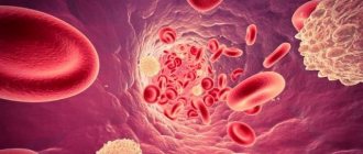
Petechial hemorrhages appear as a result of damage to small blood vessels - capillaries. When the capillaries burst, blood begins to leak under the skin.
Experts identify a huge number of reasons that can provoke the appearance of petechial hemorrhages on the skin of an adult or child. These include the following:
- traumatic injury to the skin or local injury;
- sunburn;
- allergies to certain insect bites;
- autoimmune type pathologies;
- viral and bacterial infections;
- radiation, chemotherapy – methods of treating cancer;
- the level of platelets in the blood is significantly lower than normal;
- bone marrow cancer, leukemia. These diseases significantly reduce the number of platelets in a person's blood;
- severe vomiting and dehydration - more common in newborns;
- strenuous physical activity. For example, lifting weights, labor;
- sepsis;
- vasculitis;
- scurvy;
- fevers of viral origin - Ebola, dengue fever, yellow fever cause the blood to clot poorly and bleeding to occur under the skin.
Petechial rashes can also occur as a result of taking certain medications. Medicines that may cause rash as a side effect include the following:
- antibiotics;
- anti-depression and sedatives;
- hormonal contraceptives;
- blood thinners;
- medications that help regulate your heart rhythm;
- non-steroidal anti-inflammatory drugs - NSAIDs;
- sedative medications.
If, after taking certain medications, you notice the formation of a petechial rash on the face and other parts of the body, then you should immediately consult with your doctor and select other medications.
Pathogenesis
As a result of traumatic capillary injury, platelets clump together to help the blood clot. Groups of platelets react with factors of the blood coagulation system, blood stagnates in the affected area, and a blood clot forms. In people with existing disorders in the body, the blood coagulation system works less efficiently, and small bruises - petechiae - appear on the skin.
- Primary petechiae are small dots that are initially purple or bluish-black in color and later change color to brown or yellow-brown. This is explained by the formation of hemosiderin in tissues. Gradually, the outlines of the petechiae become blurred and their color fades.
- The mechanism of formation of secondary petechiae is the leakage of blood cells into adjacent tissues. Such hemorrhages do not go away on their own. Patients require surgical intervention.
Some infectious diseases are manifested by the appearance of petechiae on the skin.
This is especially true for children with a weak immune system. In a child, the infectious syndrome is manifested by fever, tachycardia, and shortness of breath. Pathogenic microorganisms produce toxins that damage the walls of blood vessels. Subcutaneous hemorrhages or hemorrhagic rash are a characteristic sign of sepsis. In severe cases, the rash quickly spreads throughout the body, causing fainting, convulsions and delirium.
In systemic diseases, the body’s own vessels are perceived as foreign.
The immune system produces antibodies, immune complexes are formed, which circulate in the blood, settle on the walls of blood vessels and infect them. Patients develop general and specific symptoms: shortness of breath, hyperhidrosis, cardialgia, muscle and joint pain.
If petechiae are accompanied by malaise and fever, spread throughout the body, become large, and look like bruises, you should immediately consult a doctor.
Varieties
Depending on what exactly triggered the development of petechiae, they may differ from each other. Experts identify several main types of pathology.
- Vasculitis and autoimmune pathologies. In this case, petechiae form on the lower and upper extremities. After recovery, such petechiae disappear, and the skin at the site of their formation begins to peel off greatly.
- If the disease is provoked by staphylococcus, then petechial hemorrhages are observed on the mucous membrane of the hard palate (there is a photo of the examination in this article) and the skin.
- Due to gonorrhea, petechiae affect the lower part of the legs. At the same time, other symptoms of gonorrhea are clearly expressed.
- Enterovirus infection. In this case, petechiae indicate a person’s recovery. They form on the back, chest and face. They disappear within a couple of days, and not a trace remains after them.
- Meningitis. Petechiae look like a hemorrhagic rash that very quickly covers the entire body. Most of the formations occur on the buttocks, legs and abdomen of the patient.
The main types of petechiae in various diseases
Meningitis
Meningitis manifests itself as a hemorrhagic rash, the elements of which are star-shaped and pale in color. This is an early symptom of the disease, appearing in the first hours and days and spreading very quickly throughout the body. Petechiae are localized on the thighs, legs, buttocks, feet, and lower abdomen. They have a vesicle in the center and often merge with each other, forming extensive ecchymoses, which subsequently often undergo necrosis.
Gonorrhea
With gonorrhea, the rashes are localized on the distal parts of the extremities, above the large joints. They resemble pustules with hemorrhagic contents and are combined with characteristic clinical symptoms - signs of damage to the genitourinary system, anorectal area and pharynx.
Staphylococcal infection
Staphylococcal infection is manifested by purulent petechiae, upon examination of which accumulations of gram-positive cocci are detected. With staphylococcal sepsis, the permeability of vascular walls increases under the influence of microbial toxins. Hemorrhages appear in the form of pinpoint petechiae on the skin, oral mucosa and sclera.
Autoimmune diseases, vasculitis
In autoimmune diseases, petechial exanthema occurs on the arms and legs, and after 2-4 days multiple petechiae appear on it. Their appearance is accompanied by signs of intoxication syndrome: fever, malgia, arthralgia, malaise. Petechiae disappear after a few days, and in their place pigmented areas and areas of peeling remain.
hemorrhages with various vasculitis
Petechial rash with hemorrhagic vasculitis is accompanied by joint damage and abdominal pain. Most often, the large joints of the legs become inflamed - the ankle or knee. Epigastric pain is moderate in nature without obvious signs of dyspepsia. In severe cases, sudden, paroxysmal, colic-like abdominal pain is accompanied by diarrhea, vomiting, and fever.
Schamberg's disease
Schamberg's disease is hemosiderosis of the skin, resulting from autoimmune inflammation of the skin capillaries. Small dots appear on the skin of patients, as if pricked by a needle. At first they have a brown or brown color, and then lighten and disappear for a while. The rashes are located symmetrically on the body, but they are morphologically diverse. This is due to the simultaneous appearance of fresh and old elements on the skin. This disease has a benign course, since only the skin capillaries are affected. Men are more susceptible to pathology. Petechiae are located on the thighs and legs, have different sizes and uneven contours.
Enterovirus infection
Enterovirus infection is manifested by fever, muscle pain, inflammation of the soft meninges, herpetic sore throat, and gastrointestinal dysfunction. After the appearance of a petechial rash on the skin, the condition of the patients noticeably improves, and the body temperature returns to normal. The rash appears within one day. It is located on the face and torso and disappears without a trace by the end of the second day.
Diagnosis of diseases manifested by petechial rash includes collecting complaints, interviewing the patient, conducting laboratory tests and diagnostic tests:
- Coagulogram,
- General blood analysis,
- Bone marrow biopsy.
Signs of pathology
The only sign of the disease is the appearance of a petechial rash on the skin (you can see the photo in this article). Along with the rash, some other symptoms of the pathological condition may occur:
- hematomas;
- gums begin to bleed;
- nosebleeds begin;
- critical days are very difficult;
- hemorrhage occurs in the joint cavity.
Petechiae in children
In most cases, petechiae appear in children as a result of various injuries. Children prefer active games, so bruises and abrasions are their constant companions and are considered quite normal.
Subcutaneous hemorrhages can also develop in the child’s mouth. They are located on the mucous membrane and palate. The cause is food that is too hard for the child, which has severely damaged the oral mucosa. In addition, the formation of a large number of petechial-spotted rashes can be provoked by insufficient nutrition, a lack of vitamin K in the child’s body, or childhood scurvy.
Another common cause is septicemia. In most cases, this cause manifests itself in very young children who have a weak, incompletely formed immune system. It cannot completely kill all pathogenic microflora. Septicemia can occur along with other diseases. This concept refers to infection of the blood by a variety of bacteria. The skin rash will form very quickly and then spread to the entire body, which may cause the child to faint or become delirious.
Important! If you suspect septicemia in a child, you should immediately consult a doctor, because the petechial type of bleeding can be fatal. But still, in most cases, petechiae begin to appear in children due to various injuries received during play.
Etiology
Before you begin treatment for petechiae, it is necessary to identify and eliminate the causes of their appearance!
Physiological reasons
- The most common cause of petechial rash formation in healthy people is trauma—strong physical impact on the skin. The capillaries rupture and blood flows under the skin. In adults, petechiae are most often formed after a blow, and in children - during games or a fall. Hemorrhages appear on the oral mucosa when eating solid foods.
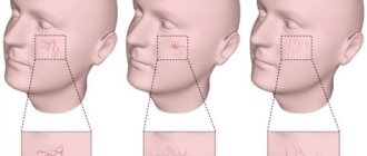
- Excessive strain, such as during bouts of coughing, emotional crying, or vomiting, can cause capillaries near the eyes and other areas of the face to break.
- Petechiae can often be seen after childbirth in a woman and newborn. Severe overexertion and stress have a negative impact on the skin of mother and child.
- Single petechiae appear during certain sports - weightlifting.
- Tight and uncomfortable clothing is the cause of petechiae.
- Petechiae may appear when the tourniquet is pulled or due to increased pressure on soft tissue. Pinpoint hemorrhages in such cases do not pose a particular health hazard and disappear without a trace after a few days.
- Skin aging.
Pathological causes
As a result of most hematological and autoimmune diseases, the formation and functioning of platelets is disrupted, which is clinically manifested by the appearance of petechiae on the skin. Disseminated intravascular coagulation is also a cause of hemorrhagic rash of bacterial origin.
- Autoimmune diseases - SLE, scleroderma, spondyloarthritis, thrombocytopenic purpura, hemorrhagic vasculitis;
examples of petichiae in thrombocytopenic purpura, characteristic of children
- Infectious diseases - endocarditis, typhoid, smallpox, sepsis, mononucleosis, scarlet fever, tonsillitis, meningitis, cytomegalovirus infection;
- Hypovitaminosis - lack of vitamins K, C;
- Capillary toxicosis;
- Hormonal dysfunction - hypercortisolism;
- Hematological disorders – thrombocytopenia and leukopenia;
- Tumors;
- Drugs;
- Long-term use of certain medications: anticoagulants - Warfarin, Heparin, Naproxen, Penicillin, Indomethacin, Atropine;
- Chemotherapy and radiation therapy;
- Children's scurvy develops with insufficient care, unbalanced nutrition and is manifested by the appearance of scattered pinpoint hemorrhages on the skin of the oral cavity.
When should you contact a specialist?
In any case, immediately after the appearance of the rash, you need to consult a specialist, because this rash can signal the development of quite serious diseases. The doctor will examine the mucous membranes and skin, after which he will be able to tell you what causes the problem and whether they can be classified as serious.
Along with the formation of petechiae, some other symptoms may appear that will indicate a serious condition of the person or child. These include the following:
- loss of consciousness or confusion;
- very high body temperature rises;
- bleeding begins;
- I constantly have a severe headache.
If any of these symptoms are present along with the rash, then you should consult a doctor immediately, because this may be a signal of the development of a very serious pathology.
Skin rashes in children: rashes, exanthemas, enanthemas
Rashes on the skin (exanthema, exanthema ) and mucous membranes (enanthema, enanthema ) can occur not only with viral and bacterial infections, but also with diseases of a non-infectious nature.
It is important to decide whether these changes represent a primary injury to the child's skin or whether the clinical signs have changed due to secondary factors (infection, trauma, or treatment). Examination by a pediatric dermatologist Moscow - Markushka clinic.
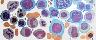
Elements of rashes in a child, children. Primary and secondary elements
There are primary and secondary elements of rashes. Primary elements are classified as roseola, spot, papule, nodule, wheal, vesicle, vesicle, hemorrhage. Secondary elements include pigmentation and depigmentation, scale, crust, erosion, crack, abrasion, ulcer, scar, cicatricial atrophy, lichenification, vegetation.
Primary elements of rashes in a child, children: roseola, spot, erythema, hemorrhage, pinpoint hemorrhages - petechiae, papule, tubercle, node, blister, vesicle, bubble
Roseola is a pale pink or red speck ranging in size from 1 to 5 mm. The shape is round or irregular, the edges are clear or blurred, does not protrude above the skin level, disappears when the skin is pressed and stretched. Roseola occurs in many infectious diseases, especially typhoid fever. Multiple roseola 1-2 mm in size are usually described as a pinpoint rash (with scarlet fever), in the process of resolution they become covered with scales or disappear without a trace.
The spot (makula) has the same color as roseola, size - from 5 to 20 mm, does not protrude above the skin level. The shape is most often incorrect. The spot disappears when pressure is applied to the skin and appears again after the pressure is removed. Multiple spots ranging in size from 5 to 10 mm are described as a small-spotted rash (for example, with rubella - a child is vaccinated against rubella at the Markushka children's clinic). Spots 10-20 mm in size form a large-spotted rash (for example, with measles, allergies - examined by a pediatric allergist in Moscow, Markushka clinic).
Erythema is large areas of hyperemic skin that are red, purple-red, or purple in color. It occurs as a result of the fusion of large spots formed by the dilation of blood vessels not only of the papillary layer of the skin, but also of the subpapillary vascular plexus. Spots larger than 20 mm that tend to coalesce should be considered erythema. Erythema is most typical for erysipelas, thermal, and ultraviolet burns .
Hemorrhage (haemorrhagia) is bleeding into the skin as a result of destruction of skin vessels. It looks like dots or spots of various sizes and shapes and does not disappear when the skin is stretched. The color is initially red, purple or violet, then, as the hemorrhage resolves, it becomes yellow-green and finally yellow (formation of hemosiderin during the breakdown of red blood cells). Color changes are clearly visible in larger hemorrhages.
Pinpoint hemorrhages are called petechiae (petechia). Multiple round hemorrhages measuring 2 to 5 mm are described as purpura . Irregular hemorrhages measuring more than 5 mm are called ecchymoses . Hemorrhages may overlap with other elements of the rash. In such cases, they speak of petechial transformation of roseolas, spots, papules. As a rule, this is observed in severe cases of the disease. Hemorrhagic rashes are detected with typhus (often in combination with roseola - roseola-petechial rash), hemorrhagic fevers, and sepsis. Hemorrhagic elements of irregular shape on a dense basis (stellate rash) are characteristic of meningococcemia and pneumococcal sepsis. Minor hemorrhages can also have a non-infectious origin (capillary toxicosis, toxic-allergic vasculitis, vitamin deficiency C, etc.).
Papule (papula) is an element of the rash that rises above the level of the skin, which is often determined by touch. It has a flat or dome-shaped surface, size - from 1 to 20 mm. The shape and color are the same as those of roseolas and spots. Papules often leave behind pigmentation and flaking of the skin. Papules that merge with each other form plaques, and when the latter merge, areas appear that are located on large areas of the skin, the size of a palm or more. Often, during a routine clinical examination of a child, it is very difficult or even completely impossible to distinguish roseola from papules. On the other hand, the same sick child can simultaneously have roseola, papules (typhoid fever, paratyphoid fever, infectious mononucleosis), papules and spots (measles - child measles vaccination, children's medical).
A tubercle (tuberkulum) is a limited, dense, cavityless formation protruding above the surface of the skin with a diameter of 1-2 to 5-10 mm. The tubercles are formed as a result of the accumulation of a specific inflammatory infiltrate in the dermis. Clinically, the tubercle is similar to a papule, but differs from it in that when palpating the tubercle, a dense infiltrate in the skin is always clearly visible. In addition, tubercles, unlike papules, undergo necrosis during reverse development, often form ulcers and leave behind a scar or cicatricial atrophy of the skin. The tubercles are most typical of cutaneous leishmaniasis, leprosy and tuberculous skin lesions, tertiary and late congenital syphilis.
A node (nodus) is a cavityless, limited compaction that goes deep into the skin, often standing above the skin level. The size of the knots ranges from a hazelnut to a chicken egg and more. They are formed as a result of the accumulation of cellular infiltrate in the subcutaneous tissue and the dermis itself. Inflammatory nodules have a soft or doughy consistency, unclear boundaries, and the skin over them is red. Nodules that appear as a result of specific inflammation (colliquatic tuberculosis, syphilitic gumma) have a dense consistency, are sharply demarcated from the surrounding tissues, and are prone to decay and ulceration with subsequent scarring.
A blister (urtica) is an acutely inflammatory, cavity-free element slightly elevated above the skin level, measuring from 2-3 to 10-15 cm or more, has a round or oval shape, and is often accompanied by itching. Color - from white to pale pink or light red. The blister usually forms quickly and disappears quickly, leaving no trace behind. It occurs as a result of limited acute inflammatory swelling of the papillary layer of the skin and simultaneous expansion of the capillaries. The appearance of urticarial elements is characteristic of allergic reactions of various origins (drug, food, cold allergies), including those of an infectious nature. Sometimes occurs in the pre-icteric period of hepatitis B (vaccination of a child against hepatitis in the Markushka children's clinic).
A vesicle (vesicula) is a cavity element measuring from 1 to 5 mm, representing a detachment of the epidermis. Usually the bubbles are filled with transparent, cloudy or bloody contents, they can shrink and give a transparent or brown crust. If the cover of the bubble is opened, then erosion is formed - a wet surface of pink or red color limited by the size of the bubble. The bubbles do not leave any scars on the skin. If a large number of leukocytes accumulate in a vesicle, it turns into an abscess - a pustule. Inflammatory changes are noted at the base and around the vesicle. Pustules are divided into single-chamber (chickenpox) and multi-chamber (natural smallpox). A group of blisters located on inflamed skin is called herpes. Vesicles are characteristic of herpes and enterovirus infections, chickenpox and natural pox. Chickenpox vaccination - Markushka Children's Clinic.
Bubble (bulla) is a cavity element with a diameter of up to 3-5 cm, located in the upper layers of the epidermis and under the epidermis. The contents of the blisters can be serous, bloody, or purulent. They can collapse, forming a crust, or open up, forming an erosive surface that turns into unstable pigmentation. The bubble occurs more often against the background of an erythematous spot, less often - against the background of unchanged skin (neonatal pemphigoid). The elements can be located both inside the epidermis, in the styloid layer (pemphigus vulgaris), and under the epidermis (multiform exudative erythema, dermatosis herpetiformis). It is observed with bullous form of erysipelas, sometimes with chicken pox, thermal burns.
Secondary elements of rashes in a child, children: hyperpigmentation, depigmentation, scales, erosion, abrasion, ulcer, cracks, tears, crust, scar, lichenification, vegetation
Secondary morphological elements are formed as a result of the evolution of the primary elements of the rash .
Hyperpigmentation (hyperpigmentatio) is a change in skin color as a result of an increase in melanin in it or the deposition of hemosiderin of primary elements.
Depigmentation (depigmentatio) occurs as a result of a decrease in the melanin content in the skin, observed after the disappearance of a nodule, tubercle - resolution of spotty-flaky (pityriasis versicolor, eczematoids) and papular (psoriasis) elements.
Scale (sguama) is an accumulation of rejected cells of the stratum corneum, sometimes the underlying layers of the epidermis. Scales occur on primary morphological elements - papules (psoriasis, syphilis), tubercles, after the resolution of blisters (eczema), etc.
Erosion (erosio) is a skin defect within the epidermis as a result of the opening of a vesicle, blister, or abscess, repeating their shape and size. When vesicles and pustules merge, erosions have scalloped edges. Erosion can also occur as a result of maceration of the skin in the area of folds or during maceration of other elements of the rash, most often papules. When erosion heals, there is no scar left; usually there is only temporary pigmentation.
An abrasion (excoriatio) is a violation of the integrity of the skin that occurs as a result of scratching, scratching, or other damage. Abrasions can be superficial - within the epidermis, sometimes involving the papillary dermis, and heal without a scar. Deeper abrasions, involving the deeper layers of the dermis, leave behind a scar. Abrasions are characterized by a tendency to become infected.
An ulcer (ulcus) is a deep skin defect that reaches the dermis, subcutaneous fat, fascia, muscles, and bones. It occurs as a result of the breakdown of the tissue of the primary element (tubercle, node, ecthyma). Its size is from 1 mm to the size of a coin or palm and more; the shape can be round, oval, linear, oblong, irregular. The surrounding tissue is either inflamed (edema, hyperemia) or infiltrated. Ulcers always heal with the formation of scars.
Cracks, tears (fissura, rhagades) - linear damage to the skin in the form of its rupture, resulting from excessive dryness due to loss of elasticity due to inflammatory infiltration or overstretching of the skin. Cracks can be located within the epidermis and dermis. They are usually localized in the corners of the mouth, interdigital folds, on the palms, soles, above the joints, and in the anus. A superficial crack after healing leaves no traces. After healing of deep cracks, linear scars remain.
A crust (crusla) is formed on the skin as a result of drying of the discharge of a weeping surface (vesicle, vesicle, abscess, ulcer, erosion). The crusts can have different colors (with serous exudate, transparent with a yellowish tint; with purulent exudate, yellow, greenish or brown; with hemorrhagic exudate, brown or black) and shape (layered, oyster-like, etc.).
A scar (cicatrix) is the formation of connective tissue at the site of a deep defect. Occurs after healing of deep skin defects at the site of ulcerated tubercles, deep pustules, nodes, deep burns, wounds. Scar formation is accompanied by the death of sebaceous and sweat glands, hair follicles, blood vessels and elastic fibers, and the disappearance of the skin pattern. Typically, scars are located below the skin level or are at its level, less often they rise above the skin level - hypertrophic scars.
Lichenification (lichenificatio) is a focus of increased skin pattern, accompanied by thickening and compaction, hyperpigmentation, and dryness. Foci of lichenification are most often localized in the neck, elbow and popliteal folds, wrist and ankle joints, inguinal folds, scrotum and occur in chronic dermatoses accompanied by itching (eczema, neurodermatitis).
Vegetation (vegetatio) is a papillary thickening of the skin that occurs as a result of the growth of the styloid layer of the epidermis and papillomatosis of the dermis during a long-term inflammatory process. More often it forms in the area of papular elements and ulcers. Vegetations can erode, bleed, and are prone to secondary infection.
Treatment with medications
After determining the cause of the problem, the specialist may prescribe the following medications:
- antibiotics are prescribed to treat bacterial infections;
- to reduce the inflammatory process, it is necessary to take corticosteroids;
- if an autoimmune pathology is present, then medications such as Methotrexate, Azathioprine or Cyclophosphamide may be prescribed;
- Biological therapy or chemotherapy is used to treat cancer.
If petechiae began to develop not due to the appearance of some disease, then rest, drinking plenty of warm liquid and special means to eliminate painful sensations would be an excellent therapy. Your doctor may prescribe pain relievers such as Tylenol, Ibuprofen, or Acetaminophen.
If subcutaneous hemorrhage occurs due to injury, there is no need to worry, because this does not pose any threat to human life. In this case, the rash must be treated with ointments against bruises. If the problem occurs in the oral mucosa, then you should exclude solid foods from your diet, and after some time the rash will disappear on its own.
Can complications arise?
The formation of subcutaneous hemorrhages of the petechial type does not provoke the appearance of any complications. As a rule, such manifestations disappear without any trace and do not even leave scars.
But if a petechial rash occurs as a result of an underlying pathology, then certain complications may arise, for example:
- damage to internal organs;
- diseases of the cardiovascular system;
- infections begin to develop in other parts of the body.
Diagnosis and treatment
Petechiae that arise spontaneously and are not accompanied by clinical symptoms go away on their own and do not require special treatment. But despite this, people who do not have any diseases need to make sure that there are no hidden causes of hemorrhages.
- If the cause of the petechial rash is injury
, a cold compress will help. It will reduce inflammation and prevent further spread of the rash. To do this, ice is wrapped in a towel and applied to the affected area for 15 minutes. - In case of an infectious process,
patients are prescribed large doses of antibiotics, taking into account the sensitivity of the isolated microbes. Antiviral, immunostimulating, detoxification, sensitizing and symptomatic therapy is carried out. - If the petechial rash is of allergic origin
, the allergen is eliminated and desensitization is carried out. For severe itching, antihistamines are prescribed - Suprastin, Zodak, Zyrtec. - For systemic autoimmune pathologies,
desensitizing drugs, corticosteroids, and vasoconstricting drugs are prescribed - “Ascorbic acid”, “Calcium chloride”, “Rutin”. The course of treatment is long – 4-8 weeks. - General restorative therapy for weakened patients
consists of prescribing vitamins K, P, C, liver extract, transfusion of red blood cells or blood, and administration of globulins.
Preventive measures
The main method of prevention can be called avoiding the causes that can trigger the development of underlying diseases. To minimize the risk, you should follow these simple tips:
- play sports;
- avoid infection;
- observe the rules of hygiene;
- practice only protected sex;
- Avoid taking medications that can cause petechiae.
Of course, you cannot avoid all the factors that can trigger the problem, but these simple tips will help reduce the risk of developing many diseases.