Detailed description of the study
An extended blood test is a primary study that is used to assess the general condition of the body, identify a wide range of diseases and monitor their treatment.
The main indicators that are included in the extended clinical blood test:
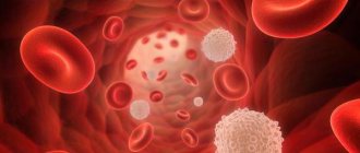
Erythrocytes are cells also called red blood cells. The main function of red blood cells is to transport oxygen to tissues and remove carbon dioxide (CO2) from them.
Hemoglobin is the main protein in red blood cells. It directly allows for gas exchange in tissues by combining with oxygen, or CO2. Insufficiency of hemoglobin in the body, along with a decrease in red blood cells, serves as the basis for the development of anemia.
Hematocrit is the ratio of all formed elements of blood (mainly red blood cells) to the liquid component of blood. Allows you to judge the excess or deficiency of red blood cells, dehydration or overhydration of the body.
The color indicator reflects how adequately the saturation of erythrocytes with hemoglobin is, helps the specialist to establish anemia and differentiate it into normochromic, hypochromic or hyperchromic.
MCH (Mean Cell Hemoglobin) - an indicator reflecting the average hemoglobin content in red blood cells, is a modern and more accurate analogue of the color indicator.
MCV (Mean Cell volume) determines the average volume of a red blood cell. Used in conjunction with MCH to identify anemia and its causes.
RDW (Red cell Distribution Width) reflects the heterogeneity of red blood cells by volume. The indicator is also important in the diagnosis of anemia; it depends on the mean erythrocyte volume (MCV), therefore, when MCV increases or decreases, its increase is observed, which indicates that there are cells of different volumes in the blood.
MCHC (Mean Cell Hemoglobin Concentration) helps estimate the mean hemoglobin concentration in red blood cells. Changes in the indicator are observed in cases of impaired hemoglobin synthesis, hypochromic and hyperchromic anemia.
Platelets also belong to the formed elements of blood; they play a key role in the processes of hemostasis and ensure timely blood clotting in case of vascular damage. Changes in this indicator may be associated with impaired cell production in the bone marrow or their accelerated destruction.
Leukocytes are called white blood cells. They participate in various immune processes and protect the body from infection. There are several types of leukocytes: eosinophils, neutrophils, lymphocytes, basophils, monocytes. Their percentage ratio allows you to evaluate the leukocyte formula to understand the current state of the immune system, identify and determine the severity of inflammatory and allergic processes in the body.
Neutrophils , the largest population of white blood cells, are aimed at fighting foreign microorganisms - including bacterial infections - in the body.
Lymphocytes play an important role in recognizing pathogenic microorganisms and forming and regulating immunity to various infections by releasing antibodies and destroying infected cells.
Monocytes are white blood cells that can differentiate into macrophages and dendritic cells after they migrate to peripheral tissues. Viral and bacterial particles are absorbed, then target proteins appear on the surface of the cell to recognize the infectious agent by the immune system. This helps in fighting pathogens and building adaptive immunity. Monocytes also take part in cleansing the body of dead cells and tissue healing.
Basophils and eosinophils are actively involved in the development of allergic reactions. They contain many granules with bioactive molecules that cause allergy symptoms. Eosinophils also play a role in protecting the body from parasites.
Reticulocytes serve as precursors of red blood cells. Their number reflects erythropoiesis - the rate of formation of red blood cells in the bone marrow. This indicator is important when assessing the effectiveness of anemia therapy. Additionally, the percentage of reticulocytes, the content of their immature forms and the production index of these cells are determined. Taken together, these data are used to manage patients with hematopoietic disorders.
Taken together, the indicators of an extended clinical blood test are necessary for the doctor as a stage of primary laboratory diagnosis of a wide range of diseases. The test can also be used to assess general health.
Blue blood
As for aristocrats, the expression “blue bloods” arose due to the paleness of their skin. Until the twentieth century, tanning was not in fashion, and the aristocrats themselves, especially women, hid from the sun, which protected their skin from premature aging and looked appropriate for their status, that is, they differed from the serfs who “plowed” all day in the sun. We now understand that pale skin color with a blue tint is actually a sign of less health.
But scientists also claim that there are about 7,000 people in the world whose blood has a blue tint. They are called kyanetics (from the Latin cyanea - blue). The reason for this is not the same hemoglobin. Their protein contains more copper than iron, which during oxidation acquires a blue tint instead of the red we are accustomed to. These people are considered more resistant to many diseases and even injuries, as their blood is said to clot several times faster and are not susceptible to many infections. In addition, there are different theories about the origin of kianeticians, including that they are descendants of aliens. There is not much information about them on the Internet, but there are articles in foreign publications where the birth of such children is explained by the abuse of rudimentary drugs long before conception. As they say, “Don’t smoke, girl, the children will be green!”, but the results from birth control may turn out blue (meaning the color of blood).
References
- Nazarenko, G.I., Kishkun, A.A. Clinical evaluation of laboratory results. - M.: Medicine, 2006. - 544 p.
- Tatkov, O.V., Stupin, F.P. General blood analysis. Information collection. - M.: Publishing solutions, 2021. - 72 p.
- Fauci, A., Braunwald, E., Kasper, D. et al. Harrison's principles of internal medicine, 17th edition, 2009.
- Pagana, K., Pagana, T., Pagana, Th. Mosby's Diagnostic and Laboratory Test Reference. 14th Edition. Elsevier Publishing Hall, 2019. - 1094 p.
Brown discharge in women
Often in the practice of a gynecologist, diseases of the female reproductive system are encountered, which are accompanied by pathological discharge.
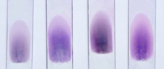
A woman should be very careful about brown discharge. Such discharge indicates the presence of bloody or bloody additions to it, which is a disorder in the female genital area.
Dark brown discharge is associated with a large amount of blood being released, while a light brown tint means less blood is being released. Discharge of scarlet blood indicates the presence of fresh bleeding (for example, due to injury to the vaginal mucosa during sexual intercourse). The blood does not have time to clot, so it has a scarlet color. Brown discharge is clotted blood.
Blood takes longer to leave the uterus when there is little of it, so it oxidizes in the process, darkening and acquiring a shade of brown and a glandular specific odor. When released, the blood mixes with vaginal lubrication and looks like brown mucus or smear.
Important! If you suddenly notice brown discharge in your vagina, then mark it on your menstrual calendar and pay attention to other symptoms.
Causes of vaginal bleeding
Postpartum period
After separation of the placenta, the uterus is a wound surface, so bleeding is normally observed. In the first days after childbirth, there may be copious brown discharge from the vagina; postpartum women are forced to use special absorbent pads designed for the postpartum period. Gradually, the amount of discharge decreases, it becomes bloody, and by 2-3 weeks it is replaced by leucorrhoea. Prolonged uterine bleeding, discharge with an unpleasant or fetid odor, or streaks of pus should be reported to the treating obstetrician-gynecologist.
Inflammation of the genital organs
A common cause of light brown or “meat slop” colored discharge is inflammatory diseases of various parts of the female reproductive system. Bleeding is associated with erosions and destructive changes in the epithelial layer, damage to small vessels. These diseases are characterized by a combination of brown discharge with other leucorrhoea (cloudy, mucous with a specific odor, purulent). Bloody discharge begins against the background of pain in the lower abdomen and increased body temperature. The symptom is caused by:
- Damage to the uterus
: endometritis, cervical erosion, endocervicitis. - Inflammation of the appendages:
salpingitis, oophoritis, adnexitis. - Vaginal diseases
: colpitis (vaginitis), vulvitis.
Pathologies of pregnancy
Bloody vaginal discharge is one of the reliable signs of spontaneous abortion or late miscarriage. The appearance of sanguineous discharge and blood is preceded by discomfort above the pubis, which is gradually replaced by a nagging, aching pain and radiates to the sacrum. Spotting brownish and then bloody discharge usually first bothers you periodically, then if left untreated it becomes constant and can turn into massive bleeding. In addition to abortion, bleeding from the genitals can be caused by:
- Ectopic pregnancy.
At first, women report spotting brown discharge that occurs due to the cessation of menstruation and aching pain in the lower abdomen. Termination of a tubal or cervical pregnancy is accompanied by massive bleeding with clots. - Chorionic carcinoma.
Bloody discharge in 80% of patients begins shortly after birth. When a tumor node is infected, the discharge is purulent and mixed with blood. Bleeding 3-5 weeks after a miscarriage or abortion is typical for a placental polyp. - Birth injuries.
The discharge of bright red blood is observed when the genital tract ruptures during the passage of the fetal head. The complication is more common during rapid labor or in the case of a clinically narrow pelvis. Massive blood loss is accompanied by shock. - Placental abruption.
Taking into account the age of detachment, the blood may have a scarlet or dark cherry color. In this case, the woman feels severe abdominal pain radiating to the perineum. In severe cases of abruption, the condition may be complicated by the development of Couveler's uterus.
Injuries
The flow of bright scarlet blood from the vagina after falls or bruises of the perineum is caused by damage and ruptures of the genital organs. The symptom is accompanied by sharp pain in the groin, difficulty urinating, and sometimes blood is found in the urine. Bloody discharge occurs with injuries to the genital organs in girls associated with household or sports injuries to the groin area. In teenage girls, heavy bleeding occurs during voluntary or violent sexual activity. Vaginal bleeding can be caused by reasons such as:
- Violent sexual intercourse
. Minor bleeding is observed due to trauma to the vaginal mucosa. Such bleeding is short-lived and is accompanied by nagging pain in the perineum. - Vaginal lacerations.
There is bleeding from the genital fissure, which is combined with swelling and cyanosis of the skin of the labia majora, and sharp pelvic pain. Massive blood loss is typical for damage to the clitoris. - Perforation of the uterus.
Perforating damage to the organ associated with curettage or criminal abortion causes profuse, bloody, bright red vaginal discharge. Bleeding develops against the background of the woman’s severe general condition and severe abdominal pain.
Taking oral contraceptives
In 30-40% of women taking hormonal contraceptives, during the first 3 months after the prescription of contraceptives, scanty bleeding from the vagina is observed, usually without any odor. The reasons for their appearance are associated with the body’s adaptation to the supply of hormones from the outside, a change in the synthesis of its own estrogens. Brownish discharge is also noted when the pill regimen is not followed. Heavy bleeding indicates atrophic processes in the uterus due to hormonal imbalances and requires an immediate visit to the doctor.
Endometriosis
Periodic, odorless, spotting brownish discharge occurs in 50-60% of patients with endometriotic growths. Bleeding from the vagina occurs due to hyperplasia of endometrial tissue and damage to the uterine cervix. Scanty bloody discharge appears a couple of days before menstruation, accompanied by intense pelvic pain and discomfort during sexual relations. With diffuse endometriosis, copious bright red discharge is possible. Retrocervical endometriosis is characterized by a combination of vaginal and rectal bleeding.
Benign neoplasms
Most often, patients with uterine fibroids report brown, odorless discharge. This benign neoplasm is characterized by heavy bleeding in the middle of the cycle, which is combined with general symptoms - pain in the lower abdomen, weakness, dizziness. Bloody discharge that occurs against the background of menstrual irregularities is characteristic of atypical endometrial hyperplasia. Spotting brown discharge, mainly after sexual intercourse, is observed with polyps of the uterus or cervical canal. Scanty bleeding occurs with condylomas of the uterine cervix.
Malignant tumors
Oncological diseases are characterized by copious brown discharge with a foul odor, in which individual clots and tissue fragments are visible. With cancer of the vulva and vagina, a triad of symptoms is observed: spotting, periodic mucous leucorrhoea and pain in the perineum. For cervical cancer, moderate bleeding is typical, observed after sexual intercourse, douching, and vaginal examination. Brown discharge also appears with adenocarcinoma and sarcoma of the uterus, germ cell tumors.
Rare causes
- Complicated course of genital infections
: chlamydia, gonorrhea, trichomoniasis. - Installation of intrauterine devices
. - Iatrogenic factors
: trauma to the vaginal epithelium or endometrium during diagnostic procedures, consequences of curettage (RDV). - Emergency contraception.
- Sexual crisis in newborns.
- Pathology of the blood system
: thrombocytopenia, coagulopathy, vasopathy.
Treatment
Help before diagnosis
The appearance of bloody vaginal discharge that is not associated with uterine involution in the postpartum period or natural menstrual bleeding is an indication for immediate medical attention. Attempts to independently treat the disorder using traditional methods or medications often lead to serious complications in the genital area and other organs. If brown discharge is accompanied by pain, you should not use analgesics from the NSAID group, which increase bleeding.
Conservative therapy
Medical tactics, first of all, depend on the degree of blood loss and the cause of the formation of reddish or brown discharge. For minor brown or bloody discharge, etiotropic therapy for the underlying pathology is indicated; massive blood loss requires the use of specific hemostatic agents. Physiotherapy methods are not recommended. To treat patients with complaints of vaginal bleeding, the following are used:
- Coagulants
. The medications are designed to stimulate the formation of blood clots and quickly stop bleeding. They are prescribed only for heavy bleeding under the control of coagulogram parameters. - Hormonal drugs
. Bloody discharge caused by impaired ovarian estrogen secretion requires specific therapy. Estrogen and progesterone agents are used. - Anti-inflammatory drugs
. Reduce the amount of inflammatory mediators. Effectively reduce pain and promote healing of defects in the epithelium of the genital tract. - Antibacterial agents
. Indicated for foul-smelling discharge and signs of infection. Depending on the reasons, etiotropic medications are used: antibiotics, antiprotozoal, antifungal.
Surgery
The ineffectiveness of conservative therapy for extensive endometriosis is an indication for excision of endometrial growths followed by cauterization. For ovarian endometriosis and the formation of “chocolate” cysts, surgical laparoscopy and oophorectomy are indicated. For cervical erosions, cauterization is performed using laser coagulation. If polyps of the uterus or cervix are detected, they must be removed. To reduce trauma, endoscopic excision of the polyp is used.
In order to stop uterine bleeding, the main feeding arteries are ligated; if this method is ineffective, the uterus is amputated. Surgical interventions are performed for large benign fibroids to prevent malignancy or profuse bleeding. The method of choice is enucleation of pathological formations with preservation of the organ and restoration of reproductive function. In case of malignant lesion, extirpation of the uterus and appendages may be required, which is combined with antitumor treatment (radiation therapy and chemotherapy).
Diagnostics
A gynecologist is involved in identifying the cause of the development of bloody discharge from the vagina. The woman requires a comprehensive examination using physical and instrumental methods. The diagnostic search is aimed at establishing the root cause of the symptom and assessing general disorders in the body caused by blood loss. The most informative are:
- Gynecological examination.
The study of the mucous membrane of the vagina and cervix with vaginal speculum is necessary to identify pathological changes, erosions, signs of inflammatory processes or endometriosis. More valuable methods are colposcopy, cervicoscopy and hysteroscopy, which make it possible to detect microscopic foci of epithelial change. - Ultrasonography
. Using an ultrasound of the pelvic organs, the uterus and appendages are visualized, voluminous neoplasms, signs of diffuse growth of the endometrium, and other causes that cause odorless or odorless bloody discharge are detected. In difficult situations, when the doctor cannot verify the diagnosis, they resort to diagnostic laparoscopy. - Visualization methods
. Tumor growths can be visualized by contrasting the genital tract - hysterosalpingography. To clarify the diagnosis and assess the degree of infiltrative growth of tumors, modern research methods are used - CT and MRI of the pelvic organs. - Biopsy.
Identification of altered areas of tissue during instrumental visualization serves as an indication for collecting endometrial and uterine cervical material. Microscopic examination of cells assesses the degree of atypia, the presence of pathological inclusions or signs of malignant growth. Immunochemical analysis of materials obtained during the biopsy is also carried out. - Diagnostic curettage
. A total scraping of the mucous membrane of the uterus and cervical canal is a more informative method than a conventional biopsy, since it allows one to obtain a larger amount of material for histological analysis. Separate diagnostic curettage (SDC) is also prescribed for therapeutic purposes. - Laboratory research
. To clarify the cause of sanguineous vaginal discharge with an unpleasant odor, it is recommended to analyze a vaginal smear for microflora and culture it on nutrient media. Be sure to evaluate the female hormonal profile. General and biochemical blood tests make it possible to clarify the degree of posthemorrhagic anemia and metabolic changes.
Consultation with a gynecologist for bloody vaginal discharge
Periods of brown discharge in women and their causes
Before and after menstruation
Usually the bleeding at the beginning and at the end of menstruation is light. During these periods, the blood has a brown or even black tint because it is oxidized. Such discharge these days (before and after menstruation) is absolutely normal.
In the middle of the cycle
Some women experience spotting during ovulation. However, the volume of such secretions is small, and they can be either brown or pinkish in color. In the middle of the cycle, light spotting most often occurs in women who take oral contraceptives and who are already approaching menopause in age. In these cases, it is necessary to visit a doctor to rule out the presence of pathologies.
Instead of menstruation
If your cycle is a little off (a difference of up to 7 days is absolutely normal). But if your period does not come, you should consult a gynecologist.
During pregnancy
Brown discharge may be a sign of pregnancy. Light bleeding may occur 10-14 days after fertilization, when the egg attaches to the wall of the uterus.
Pay attention to other signs of pregnancy. These may include tender and painful breasts, nausea, frequent urination, fatigue, and vomiting.
Take a home test if you think you might be pregnant but your period is brown or late. Visit your gynecologist if the test is positive to confirm the result and plan next steps.
When taking oral contraceptives (OC)
Taking OCs affects the level of sex hormones, so brown discharge in women is most often normal in this case, especially in the first months of taking it. If such discharge is not accompanied by pain and has no odor, but continues longer than 3 months after starting to take OCs, a consultation with a gynecologist is necessary.
Causes of anemia
Anemia is always a secondary disease, that is, complications of other diseases. The main, “primary” diseases in relation to anemia can be a variety of diseases - both diseases related to the hematopoietic organs and diseases of other organs and systems. Of the blood diseases accompanied by the development of anemia, we must mention, first of all, acute and chronic leukemia and diseases leading to increased bleeding (hemophilia, thrombocytopenia). Among the causes of anemia that are not related to the process of hematopoiesis, the most prominent are diseases accompanied by acute or chronic blood loss (peptic ulcer, hemorrhoids), impaired absorption of nutrients (diseases of the stomach, intestines, pancreas), lack of certain substances in food (various vitamin deficiencies), the formation of antibodies to the cells of one’s own body (systemic lupus erythematosus, rheumatoid arthritis), various poisonings, radiation damage...