How is an electrocardiogram taken?
ECG recording is performed in a special room, maximally shielded from various electrical interference. The patient sits comfortably on the couch with a pillow under his head.
To take an ECG, electrodes are applied (4 on the limbs and 6 on the chest). An electrocardiogram is recorded during quiet breathing.
In this case, the frequency and regularity of heart contractions, the position of the electrical axis of the heart and some other parameters are recorded. This simple method allows you to determine whether there are abnormalities in the functioning of the organ, and, if necessary, refer the patient for a consultation with a cardiologist.
The concept of EOS norm
The position of the EOS depends on:
- The speed and correctness of impulse movement through the cardiac systems.
- Quality of myocardial contractions.
- Conditions and pathologies of organs that affect the functionality of the heart.
- Heart condition.
For a person who does not suffer from serious diseases, the axis is characteristic:
- Vertical.
- Horizontal.
- Intermediate
- Normal.
The normal position of the EOS is located according to Died at coordinates 0 – 90º. For most people, the vector passes the limit of 30 - 70º and is directed to the left and down.
- What problems will the electrical axis of the heart tell you about?
In an intermediate position, the vector ranges from 15 to 60 degrees.
According to the ECG, the specialist sees that the positive waves are longer in the second, aVF and aVL leads.
Alpha Angle Definition
You can also determine the position of the electrical axis using a pencil. This method is not accurate enough, and is used in many cases by students.
After this, the sharp side of the pencil is directed to the R-wave, to the lead where it is largest.
| Normal position | Determined if the sharp end of the pencil is in the lower left corner, and its reverse side is in the upper right corner |
| Horizontal | With the pencil positioned almost horizontally |
| Shift left | Determined when the pencil is practically positioned horizontally, it is impossible to accurately determine |
| Offset right | The location of the pencil is determined closer to the vertical position |
The disease is not diagnosed based on the displacement of the electrical axis of the heart alone. This factor is one of the parameters on the basis of which abnormalities in the body can be diagnosed.
These include:
- Insufficient blood supply to the heart;
- Primary damage to the heart muscle, not associated with inflammatory, tumor, ischemic lesions;
- Heart failure;
- Heart defects.
Normal position of the EOS
The EOS angle is considered one of the main parameters that is studied when deciphering ECG indicators. For a cardiologist, this parameter is an important diagnostic indicator, the abnormal value of which clearly signals various disorders and pathologies.
By studying the patient's ECG, the diagnostician can determine the position of the EOS by examining the waves of the QRS complex, which show the work of the ventricles on the graph.
An increased amplitude of the R wave in the I or III chest leads of the graph indicates that the electrical axis of the heart is deviated to the left or right, respectively.
In the normal position of the EOS, the greatest amplitude of the R wave will be observed in the II chest lead.
- Features of conducting and interpreting ECG during pregnancy
2When does the heart axis “go to the left”?
The conclusions of a functional diagnostics doctor about the deviation of the cardiac axis to the left are not an independent diagnosis. But they always give reason to wonder why the heart axis “went to the left.”
A slight shift of the EOS to -190, as well as its semi-vertical position, in some cases is not considered a pathology. This position of the axis can be observed in healthy, tall, thin people, in athletes with a trained heart, in children with an asthenic physique, and with a high position of the diaphragm dome.
If the cardiac axis is significantly deviated to the left, then this pathological condition indicates problems with the heart; the cause of such a displacement must be established. After all, this symptom can sometimes be the first “bell” in case of pathology of the heart and blood vessels.
According to some data, a deviation of the electrical axis of the heart to the left up to -29-300 is sometimes called a slight deviation, and if the angle is from -450 to -900 they speak of a sharp deviation.
Diseases associated with displacement of the OES
Deviation of the electrical position is not an independent pathology. If such a violation is observed, but there are no other pathological symptoms, this phenomenon is not perceived as a pathology. In the presence of other symptoms of cardiovascular diseases, in particular lesions of the conduction system, a shift in the EOS may indicate a disease.
Possible diseases:
- Ventricular hypertrophy.
Marked on the left side. There is an increase in the size of the heart, which is associated with increased blood flow. It usually develops against a background of prolonged hypertension, which simultaneously increases vascular resistance. Hypertrophy can also be triggered by ischemic processes or heart failure. Ventricular hypertrophy - Valve lesions. If damage to the valve apparatus develops in the area of the ventricle on the left side, axis displacement may also occur. This usually occurs due to a blockage in the blood vessels that prevents the release of blood. Such a disorder may be congenital or acquired.
- Heart block. A pathology associated with a disturbance in the rhythm of the heartbeat, which is caused by an increase in the interval between the conduction of nerve impulses. The disorder can also occur against the background of asystole - a long pause, during which the heart does not contract with further ejection of blood.
- Pulmonary hypertension. It is noted when the EOS deviates to the right side. Usually occurs against the background of chronic diseases of the respiratory system, including asthma, COPD. The long-term effect of these diseases on the lungs causes hypertrophy, which in turn provokes a change in the position of the heart.
- Hormonal disorders. Against the background of hormonal imbalance, an increase in the heart chambers may occur. This leads to disruption of nerve patency and deterioration of blood emission.
Vascular crisis: symptoms and causes of a dangerous pathology
In addition to these reasons, deviations may indicate congenital heart defects, atrial fibrillation. A shift in EOS is often observed in people who engage in intense sports or subject the body to other types of physical activity.
What influences the location of the EOS?
Before discussing the direction of the electrical axis, you should understand what the conduction system of the heart is. It is this structure that is responsible for the passage of impulses through the myocardium.
The conduction system of the heart is atypical muscle fibers that connect different parts of the organ. It begins with the sinus node, located between the mouths of the vena cava.
Next, the impulse is transmitted to the atrioventricular node, located in the lower part of the right atrium. The next to take up the baton is the His bundle, which quickly diverges into two legs - left and right.
In the ventricle, the branches of the His bundle immediately become Purkinje fibers, which penetrate the entire cardiac muscle.
An impulse entering the heart cannot escape the myocardial conduction system. This is a complex structure with fine settings, sensitively responding to the slightest changes in the body. In case of any disturbances in the conduction system, the electrical axis of the heart can change its position, which will be immediately recorded on the electrocardiogram.
What is the electrical axis of the heart?
EOS is the total (prevailing) vector of all electrical impulses that are observed in the cardiac conduction system during one contraction cycle.
Often this indicator is identical to the electrical position of the heart (EPS) - the orientation of the resulting vector of impulses from the ventricles relative to the axis of lead I on the ECG. In the myocardium, like other muscles of the body, bioelectric currents (action potentials) arise during contraction. It is their electrocardiograph that registers and records them on specialized film in the form of an ECG.
The impulse is generated by the pacemaker (sinus node), from where the excitation reaches the atrium along the nerve pathways of the heart, and then the atrioventricular node (AV). This connection inhibits transmission so that contraction follows relaxation of the atria, which ensures one-way and continuous flow of blood through the cardiac chambers.
On an ECG, electrical impulses are displayed in the form of multidirectional teeth:
- positive – P, R, T – directed upward relative to the isoline;
- negative – Q, S.
An electrocardiographic recording is a recording of changes in potential differences during the process of excitation and relaxation of the atria and ventricles, caused by the electromotive force of the heart (EMF) from the surface of the human body.
EMF is an unstable value; its direction changes throughout the entire cardiac cycle. When summing up all the instantaneous orientations of the impulses (according to the rules of addition), a vector is obtained corresponding to the average EMF during the full period of depolarization - EOS (direction of the electric motor force during the registration of QRS on the ECG).
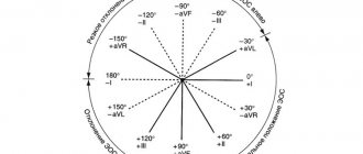
When recording an ECG, the electrodes are located in three leads that record the potential difference:
- I – left-right hand;
- II – left leg – right hand;
- III – left leg – left arm.
This placement forms a three-dimensional arrangement of EMF vectors on the torso, which forms the “Einthoven triangle”. If you put the EDS in this form, then the angle α (alpha) between the electromotive force and the horizontal of the first lead will express the deviation of the EOS.
Also, the angle α is approximately determined using the six-axis Bailey coordinate system or using special tables. In the absence of the above devices at hand, the orientation of the EOS is established by measuring the height of the R and S waves in standard leads I and III:
- RII=RI+RIII – normal axis location;
- RI>RII>RIII, SIII>RIII – left-sided deviation of the EOS;
- RIII>RI, SI>SIII – the EOS deviates to the right.
What positions of the EOS exist normally and what is the difference between them?
The muscle mass of the left ventricle (LV) is commensurately greater than the right. Therefore, the electrical processes that occur in the LV are stronger, and the EOS vector will be directed in this direction. If we project the heart onto a coordinate system, then the left ventricle will be located in the range of values +40 0 +70 0 (which is considered the normal orientation of the axis).
However, the individual structural features of the heart and the physique of each patient vary the position of the EOS within the range from 0 0 to 90 0.
Variants of the normal position of the EOS
The normal position of the EOS is the angle α from 30 0 to 69 0, the height RII≥RI>RIII, and in III and VL the R and S teeth are approximately the same. The cardiac axis is clearly perpendicular to lead III.
conclusions
Axis deviation is often not a sign of an acute condition. But if a sharp disturbance of EOS is registered, reaching a value of more than +90 0, then this may indicate a sudden disorder of conduction in the myocardium and threaten cardiac arrest. Such patients require immediate specialized medical care in order to find the cause of such a sharp change in the direction of the current.
The following sources of information were used to prepare the material.
Close in meaning to the electrical axis of the heart is the concept of “electrical position of the heart.” The electrical position of the heart means the direction of the resulting vector of excitation of the ventricles relative to axis I of the standard lead, taking it as if it were the horizon line.
There is a distinction between the vertical position of the resulting vector relative to axis I of the standard lead, calling it the vertical electrical position of the heart, and the horizontal position of the vector - the horizontal electrical position of the heart.
There is also a basic (intermediate) electrical position of the heart, semi-horizontal and semi-vertical. Figure 37 shows all the positions of the resulting vector and the corresponding electrical positions of the heart.
For these purposes, the ratio of the amplitude of the R waves of the ventricular complex in the unipolar leads aVL and aVF is analyzed, not forgetting the features of the graphic display of the resulting vector with the recording electrode (Fig. 20-23).
What options for placing an EOS can a healthy person have?
In children, the position of the EOS changes as they grow and develop
In infants under 12 months of age, the electrocardiogram shows the direction of the axis to the right. During the course of a year, children's EOS changes and becomes vertically positioned.
By the age of 2-3 years, the axis in 60% of children is vertical, in the rest it changes to normal. This occurs due to growth, enlargement of the left ventricle and reversal of the heart. In preschoolers and older children, the normal position of the EOS dominates.
Bacterial endocarditis: all about the disease and how to deal with it
Determination of the EOS (electrical axis of the heart) by ECG in normal conditions and with abnormalities – meduniver.com
The correct location of the axis in children is considered:
- Babies up to 12 months - EOS is from 90 - 170 degrees
- Children 1-3 years old – vertical direction
- Schoolchildren and teenagers note normal EOS in 60% of children
Most often, the identified deviations in EOS are a variant of the norm and arise due to the individual characteristics of human anatomy. But there are cases when the displacement is too large - this may indicate diseases, including:
- pulmonary hypertension;
- pulmonary stenosis;
- pathology of the atrial septum;
- cardiac ischemia.
Stenosis is determined on the electrocardiogram due to myocardial hypertrophy. Both congenital and acquired forms are detected. In the first case, the diagnosis can be established in early childhood when performing the first ECG.
Atrial septal defects cause a vertical position of the EOS. This happens when the hole size is sufficiently large.
With ischemia of the disease, the lumen of the coronary arteries narrows, which causes insufficient blood supply to the myocardium. In severe form, there is a risk of pathology developing into a heart attack.
During pregnancy, the EOS becomes upright quite rarely. This is due to the physiological characteristics of the body of the woman carrying the baby.
The uterus is constantly enlarging, thereby beginning to affect other internal organs. Because of this, the EOS shifts in most cases in the horizontal direction.
If the ECG showed a vertical position of the axis, the patient will require additional examination. The cause may be heart disease.
- ECG for determining EOS, interpretation of indicators, norms and deviations
In children, such placement is usually attributed to age-related characteristics. As the body matures, it acquires the proper structure, and after complete formation, the electrical axis of the heart returns to its normal location.
EOS position options
When fixing the electrical axis of the heart, the above-described positions are rarely observed. Semi-horizontal and semi-vertical axis positions are recorded in the majority of cases.
The results of the electrocardiogram may record rotations of the EOS around the coordinate axis, which may be normal. Such cases are considered individually, depending on the patient’s symptoms, condition, complaints and the results of other examinations.
Violations of the norm indicators are deviations to the left or right.
For infants, a clear axis shift is noted on the ECG; during growth, it normalizes. For the period of one year from birth, the indicator is usually located vertically.
Standards for children:
- Infants - from ninety to one hundred and seventy degrees;
- Children from one to three years old - vertical position of the axis;
- Adolescent children – normal axis position.
ECG of a newborn
ECG signs in the analysis of EOS are determined by the rightogram and the leftogram.
A rightogram is finding a vector between indicators 70-900. On electrocardiography it is demonstrated by long R waves in the QRS group. The vector of the third lead is larger than the wave of the second.
Phramogram
The levogram on an ECG is the alpha angle passing between 0-500. Electrocardiography helps to determine that the usual lead of the first QRS group is characterized by an R-type expression, but already in the third lead it has an S-type shape.
Levogram