Read information about other diseases starting with the letter “L”: Lactostasis, Laryngitis, Laryngotracheitis, Cor pulmonale, Pulmonary fibrosis, Leukemia, Leukemia in children, Leukoplakia, Leucinosis, Leprosy, Dental treatment, Lymphadenitis, Lymphangitis, Lymphogranulomatosis, Lymphoma, Lymphostasis, Lipoma, Listeriosis, Lichen, Pediculosis pubis.
Lymphangitis is a type of inflammation that affects the capillaries and trunks of the lymphatic system. The disease occurs in the presence of purulent-inflammatory processes. It is characterized by visual swelling and is accompanied by painful sensations along the location of the inflamed lymph vessels. I receive complaints from a patient about a feeling of weakness in the body and elevated temperature.
The disease can affect blood vessels of different sizes. Phlebologists and lymphologists note frequently diagnosed lymphangitis of the legs. Less commonly, the pathology affects the pulmonary lobes, mammary glands and other parts of the body. Some patients experience the development of specific venereal lymphangitis and non-venereal lymphangitis, spreading throughout the penis.
General information
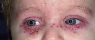
Lymphangitis (lat. lymphangoitis) is an acute inflammatory process of lymphatic vessels of various locations/calibers (lymphatic capillaries, vessels, trunks), developing as a complication of local purulent-inflammatory foci or infected skin lesions. That is, lymphangitis is a secondary disease, most often developing against the background of an infected wound , boil / carbuncle , panaritium , phlegmon , abscess , trophic ulcers , erysipelas, etc. or skin damage (microtraumas, abrasions, scratching, bites). As a rule, lymphangitis is accompanied by lymphadenitis (enlargement of the submandibular, cervical, supra/subclavian, axillary, inguinal, popliteal and other lymph nodes). The place of primary localization of the purulent-inflammatory process and lymphangitis is the upper/lower extremities (photos of lymphangitis are given below), but since the human lymphatic system is extremely extensive, the pathological process can develop on any part of the body.
Lymphangitis is accompanied by
painful swelling / hyperemia of inflamed lymphatic vessels along their course, regional lymphadenitis , edema , weakness , chills , and increased body temperature.
In andrology/urology, a condition such as “non-venereal lymphangitis of the penis” is often diagnosed. Non-venereal lymphangitis develops when the tissues of the penis are injured (during prolonged sexual intercourse/frequent masturbation). Its manifestation is the appearance of a painless, compacted (on palpation) lymphatic vessel localized along the shaft of the penis or parallel to the coronary groove. As a rule, such manifestations of lymphangitis do not require treatment, and they disappear without a trace/spontaneously within 1-2 days.
Treatment of lymphangitis
Treatment of lymphangitis is aimed at eliminating the primary focus that provoked the occurrence of the inflammatory process in the lymphatic vessels. To do this:
- treatment of existing wounds;
- opening, drainage and sanitation of felons, abscesses, phlegmons.
You cannot massage, heat inflamed tissues or rub ointments into them. It is important to provide the patient with complete motor rest.
Medications indicated:
- Antibacterial drugs (first and second generation cephalosporins, semisynthetic penicillins, lincosamides, aminoglycosides can achieve the best effect).
- Antihistamines.
- Anti-inflammatory complexes.
Physiotherapeutic procedures can include:
- laser blood irradiation (ILBI);
- ultraviolet blood irradiation (UFOI).
Surgical treatment is performed only if it is necessary to drain the abscess.
Treatment of chronic lymphangitis
In case of a sluggish chronic course of the disease, the patient is prescribed:
- semi-alcohol compresses;
- local ointment dressings;
- X-ray therapy;
- mud therapy;
- compresses with dimethyl sulfoxide;
- Ural Federal District.
Specific treatment for non-venereal lymphangitis of the penis is usually not carried out. If the pathology is caused by a sexually transmitted infection, the underlying disease is treated.
Pathogenesis
In the development of lymphangitis, a significant role is played by the anatomical and physiological specificity of lymph circulation in one or another part of the body, the localization/depth of the wound or primary inflammatory focus, the virulence of pathogenic microflora and the state of general immunity . Pathogens/their toxins initially enter the interstitial spaces, and then move into the lymphatic vessels from the primary site of infection with the lymph flow into large-caliber lymphatic vessels or adjacent lymph nodes.
Their action is manifested by a reactive inflammatory process of the walls of lymphatic vessels with swelling of the vascular endothelium, increasing its permeability, release of exudate and loss of fibrin with the formation of clots (thrombi), which contributes to the development of local lymph circulation disorders (lymphostasis). The pathological process develops as endolymphangitis / panlymphangitis with its spread towards the lymph nodes.
As the inflammatory process progresses, purulent lymphangitis , and in the absence of treatment, purulent fusion of blood clots. perilymphangitis develops with damage to muscles, blood vessels, joints, etc.
Treatment of arm lymphedema at the Innovative Vascular Center
The innovative vascular center has been developing the direction of conservative and surgical treatment of arm lymphostasis for more than 10 years, relying on the German experience of conservative treatment used by Dr. Shingale and microsurgical technology of lymphovenous anastomoses and lymph node transplantation.
We provide a full-fledged treatment and rehabilitation complex, which allows you to reduce swelling by 70-100% and control it. The course of treatment is 14 - 28 days.
Surgical treatment is used for stage 2-3 lymphedema of the arm and requires a preliminary course of conservative therapy. The experience of our clinic shows that the manifestations and progression of lymphedema can be significantly reduced and even reversed.
Classification
The classification is based on several factors, according to which they distinguish:
- Depending on the etiological factor, nonspecific and specific lymphangitis .
- Depending on the course – acute and chronic.
- According to the depth of the affected vessels - superficial and deep.
- The nature of the inflammation is serous (simple) and purulent.
- According to the caliber of the affected vessels - capillary (reticular/mesh), in which an extensive network of small capillaries is involved in the pathological process, and truncular (stem), which is based on inflammation of one/several large vessels.
Causes
The etiological factor of nonspecific lymphangitis is pathogenic pyogenic microflora. The most common causative agents of the disease include coccal flora (Staphylococcus aureus/beta-hemolytic streptococcus), Pseudomonas aeruginosa , Escherichia coli , Proteus , both in the form of a monoculture and their combinations, and in patients with immunodeficiency - anaerobes and gram-negative bacteria .
Specific lymphangitis in a patient is associated primarily with the presence of tuberculosis infection (Koch bacillus), syphilis (treponema pallidum), plague (plague bacillus), actinomycosis (actinomycete bacteria), genital herpes (herpes simplex virus type 1/2), etc.
Predisposing factors are the presence of local purulent-inflammatory foci/infected skin lesions and decreased immunity .
Stages of the disease
- Stage I: Spontaneously reversible. The swelling is quite noticeable; if you press on the skin with your finger, a hole remains. After rest (especially in the morning) it subsides, but in the evening it becomes the same. Patients rarely turn to specialists at this stage.
- Stage II: Spontaneously irreversible. The skin hardens (this is due to the growth of connective tissue), the swelling is no longer so soft and when pressing with a finger on the skin there is no hole left. The skin is very tight and sensitive. The patient may experience pain.
- Stage III: Irreversible. The skin is fibrous (the tissue scars), cysts or papillomas appear on it. The limb is deformed due to tissue damage. She moves poorly or even becomes motionless and becomes heavy. This is the extreme stage of lymphedema - elephantiasis.
Symptoms
Acute lymphangitis is characterized by severe general intoxication accompanying the purulent-inflammatory process, manifested by weakness , chills , high temperature (39-40°C), sweating and headache . With reticular lymphangitis, superficial pronounced hyperemia first appears in the area of the source of infection ( abscess , boil , etc.) with a pronounced mesh pattern against the background of increasing erythema . At the same time, reticular lymphangitis in clinical symptoms resembles erysipelas , but hyperemia has unclear boundaries uncharacteristic of erysipelas.
A local manifestation of stem lymphangitis is the presence on the skin of red stripes of narrow inflamed lymphatic vessels towards the regional lymph nodes. There is induration, swelling and pain in the inflamed cord-type cords, tension/swelling of the surrounding tissues, and often the development of regional lymphadenitis.
When deep vessels are damaged, there is no local hyperemia, but pain and swelling in the affected area quickly increases; on palpation - severe pain, lymphedema .
In the chronic course of lymphangitis, the clinical symptoms are erased and manifest mainly by persistent edema and lymphostasis caused by blockage of the deep lymphatic trunks with minimal appearance of a general nature.
Types of Postoperative Lymphedema
- Temporary reversible lymphedema: Occurs within a few days after surgery and usually lasts a short period of time. goes away quickly enough under the compression sleeve.
- Subacute lymphostasis of the arm: appears 4-6 weeks after surgery. It is characterized by pain and high tissue density, but goes away quite quickly under the influence of compression therapy.
- Chronic arm lymphedema: This painless swelling usually appears 18-24 months after surgery.
Tests and diagnostics
Diagnosis of superficial lymphangitis is not difficult and is based on clinical symptoms and the presence of a focus of primary infection. To diagnose lymphangitis of deep lymph nodes, additional laboratory and instrumental studies are carried out:
- Complete blood count (increased ESR/moderate leukocytosis).
- Blood test (bacteria culture) for sterility.
- Bacterial culture of discharge from the primary source of infection in order to identify the pathogen and determine its sensitivity to antibiotics.
- Doppler ultrasound/duplex scanning/CT. Allows you to visualize the narrowing of the lumen of the lymphatic vessels/hardening of soft tissues, the extent of the pathological process.
Differential diagnosis with deep vein thrombophlebitis , soft tissue phlegmon , osteomyelitis , erysipelas .
How is it diagnosed?
The development of regional lymphangitis is diagnosed during the examination. In order not to confuse it with superficial phlebitis, it is necessary to do laboratory tests and conduct instrumental studies.
Diagnostic signs are considered:
- the number of leukocytes in the peripheral blood is increased;
- duplex scanning and Doppler ultrasound visualize the heterogeneity of lymph vessels, a change in the lumen towards narrowing;
- CT scan determines how deep the lymphangitis has spread;
- analysis of pus allows you to determine the pathogenic microorganism.
A complicated form of the disease requires a blood test for sterility.
Prevention
Prevention consists of preventing purulent infection. A healthy lifestyle and strong immunity play a big role in this. If you have cuts or other wounds, it is important to promptly treat them with antiseptics at home. If ulcers appear ( furuncle , carbuncle , abscesses after insect bites, etc.), you need to contact a surgeon in time to sanitize the lesions and follow his instructions regarding taking antibiotics (if necessary).
List of sources
- Ermolaev V. L., Makarova N. P. Diseases of lymphatic vessels and nodes. Ekaterinburg: GBOU VPO USMU Ministry of Health of Russia, 2015. – 264 p.
- Shlyapnikov S.A. Surgical infections of soft tissues - an old problem in a new light // Infections in surgery, 2003, No. 1(1). – P. 14-21.
- Belousova T.A., Kayumova L.N., Goryachkina M.V. Systemic antibiotics in the treatment of bacterial infections of the skin and soft tissues: focus on macrolides / breast cancer. - 2011. - No. 5.
- Guchev I.A., Sidorenko S.V., Frantsuzov V.N. Rational antimicrobial chemotherapy for skin and soft tissue infections. Antibiotics and chemotherapy. 2003, vol. 48, 10, pp. 25–31.
- Surgical infection: A guide for general practitioners / Stolyarov E.A., Grachev B.D., Kolsanov A.V., Batakov E.A., Sonis A.G. - 2004.