Vascular diseases include diseases of the lymphatic vessels and veins, which are accompanied by their inflammation or blockage. A separate group of diseases includes arteriovenous malformations: this is the name for pathological and most often congenital communications between veins and arteries.
When blood vessels are damaged in the areas they feed, blood circulation is significantly impaired, and a deficiency of nutrients and oxygen occurs. This determines the severity of the disease, as well as its main manifestations.
Usually, disruption of the normal state of blood vessels is a direct consequence of an unhealthy lifestyle and many different diseases, including endocrine disorders and pathologies of the cardiovascular system. All these diseases can remain undetected for a long time, gradually progressing. As they progress, significant circulatory disorders gradually begin, which can lead to the need for surgery and even disability.
Symptoms of vascular diseases
Peripheral vascular disease has a wide variety of symptoms. However, in the initial stages of the disease they are usually nonspecific and are often attributed to physical overload or neurological disorders. This happens especially often in the case of vascular diseases of the lower extremities. When the vessels that supply the brain and internal organs are affected, this leads to serious disruptions to their functioning.
When the vessels of the extremities are damaged, the patient complains of:
- convulsions (both prolonged, which are accompanied by severe pain, and isolated spastic twitchings);
- numbness, as well as tingling of varying degrees of intensity in different places;
- weakness in the limbs;
- the skin becomes dry and thin;
- lameness that occurs periodically;
- a mesh appears on the limbs, consisting of spider veins, dilated small vessels or noticeable vascular trunks;
- the growth of nails and hair is disrupted;
- Dark brown spots, lumps, and ulcers appear on the skin.
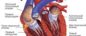
Features of the human cardiovascular system
The cardiovascular system is a branch of veins and arteries that reach the most distant parts of the body. Her work is based on her heart. This organ, through its contractions, creates pressure for the movement of blood along the vascular bed and sets a certain pace. In the human body there are two circles of blood circulation: large and small. In general, the liquid moves as follows:
- from the left ventricle of the heart, arterial blood moves through the aorta and large arteries to the capillaries;
- gas exchange occurs in the capillary network: cells receive oxygen, and the blood is saturated with carbon dioxide;
- venous blood flows through small venules, which then connect into large veins and empty into the right atrium.
The pulmonary circulation begins from the right ventricle. Through the pulmonary artery, venous blood enriched with oxygen flows to the lungs, where it is saturated with oxygen. Then it returns to the left atrium to carry the necessary substances to all organs and tissues.
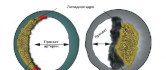
IMPORTANT! Normally, the walls of blood vessels are strong and elastic. With age or due to illness, they can lose these qualities, which is why blood does not flow to distant parts of the body. Circulation can also be impaired when sediment accumulates on the inner wall of veins and arteries.
Who is at risk?
Those who lead an unhealthy lifestyle, as well as those who are predisposed to them from birth, are most at risk of developing vascular diseases.
There are general risk factors for vascular damage, which include:
- smoking – it is one of the main risk factors; male gender;
- frequent and severe stress;
- rare physical activity;
- work that involves lifting heavy objects, standing for long periods of time, or staying in one position;
- hypothermia;
- excess weight;
- a lot of salt and fat in food;
- diabetes;
- high cholesterol;
- high blood pressure.
How to avoid complications?
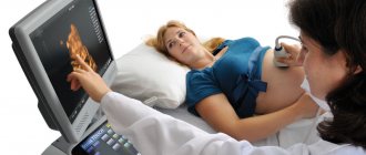
Considering that diseases affecting blood vessels do not occur overnight, but develop over a long period of time, to prevent dangerous consequences, we recommend undergoing a preventive examination. Modern methods of vascular diagnostics will help detect the disease in the early stages of its development. These include computed tomography, ultrasound, coronary angiography, etc.
In the early stages, the development of vascular diseases is stopped with the help of drug therapy and lifestyle correction. However, if these measures are not enough, endovascular interventions come to the rescue. Angioplasty and vascular stenting procedures are not inferior in their effectiveness to open surgical operations, but in comparison with the latter they are painless and do not require long-term rehabilitation. Vascular reconstruction is performed under local anesthesia, and the recovery period takes 1-2 weeks.
Diagnosis of vascular diseases
Specialists make a diagnosis based on the history of the disease, its manifestations and the results of various studies. Nowadays, Doppler-enhanced vascular ultrasound is used to diagnose vascular diseases. During laboratory tests, specialists pay special attention to the study of indicators of the blood coagulation system - coagulogram (code 138), since the most severe complications of vascular pathology entail the risk of increased thrombus formation.
Laboratory tests that are used to diagnose vascular diseases:
- clinical blood test (code 2 and code 5);
- general urine test (code 746);
- APTT (code 136);
- D-dimer (code 137);
- Fibrinogen (code 142);
- antithrombin III (code 21145);
- total protein (code 21), urea (code 19), creatinine (code 83) and potassium (code 100) in serum;
- creatinine in daily urine (code 726);
- rheumatoid factor (code 539);
- Reberg sample (code 749);
- C-reactive protein (code 26);
- Renin (code 206);
- Diagnosis of antiphospholipid syndrome (code 516 and code 517);
- Homocysteine (code 16).
Chest pain
A feeling of burning and squeezing, obvious, dull, severe or periodic pain, spasm - all these sensations in the chest are the surest sign of heart problems. With spasm of the coronary vessels, the pain is burning and acute, which is a sign of angina pectoris, which often occurs even at rest, for example at night. An attack of angina is a harbinger of myocardial infarction and coronary heart disease (CHD).
Severe, prolonged pain in the chest, radiating to the left arm, neck and back, is characteristic of a developing myocardial infarction. Chest pain during myocardial infarction can be extremely severe, including loss of consciousness. By the way, one of the most common causes of heart attack is atherosclerosis of the coronary vessels.
Chest pain radiating to the back of the head, back, or groin area is a symptom of an aortic aneurysm or dissection.
Dull and wave-like pain in the heart area, which does not spread to other areas of the body, accompanied by an increase in temperature, indicates the development of pericarditis.
However, acute chest pain may also indicate other diseases, for example, be a symptom of intercostal neuralgia, herpes zoster, sciatica in the neck or chest, spontaneous pneumothorax, or esophageal spasm.
Treatment of vascular diseases
Specialists treat peripheral vascular diseases based on the severity of their clinical manifestations and the type of damage (blockage, inflammation). During the consultation, the doctor will necessarily include in the treatment plan recommendations on proper nutrition and a healthy lifestyle, which will allow you to slow down the development of many different diseases in the early stages.
Treatment of vascular diseases includes:
- prescribing medications to the patient. Based on the examination results, the doctor selects one of the specific treatment regimens, sets the dose of the drugs and determines the duration of their use. The following medications can be used in the treatment of vascular diseases:
- medications that affect cholesterol levels;
- anti-inflammatory drugs;
- drugs that improve tissue metabolism and microcirculation, antispasmodics;
- drugs with antiarrhythmic function that lower blood pressure;
- surgical methods of treatment.
Methods for diagnosing heart disease
In addition to complaints, the doctor evaluates how the disease of the cardiovascular system developed and whether relatives have similar symptoms. Examines the patient:
- measures blood pressure;
- pulse;
- listens to heart sounds and murmurs at rest and after exercise;
- orders an ECG to assess heart rhythm;
- X-rays, MRI, EchoCG show the position of the heart, the thickness of its walls, and allow one to evaluate its contractility.
In identifying heart diseases, an important role is played by load tests - on a bicycle ergometer. They allow you to detect symptoms that do not yet appear in everyday life. At the same time, the doctor selects the required load so as not to harm the body and not cause a deterioration in the condition.
In case of rhythm disturbances and pressure changes, daily monitoring is prescribed. A person performs his usual work, sleeps, eats, while the device records ECG and blood pressure indicators. After analyzing the data, the doctor identifies those points that provoke the appearance of symptoms.
To diagnose the condition of blood vessels, contrast angiography and rheovasography are prescribed.
Prevention of vascular diseases
Prevention of vascular diseases is the reduction or complete elimination of risk factors, as well as the normalization of blood flow.
This is mainly facilitated by a healthy lifestyle:
- weight control;
- avoiding hypothermia;
- proper nutrition, which limits the total caloric content of food, as well as the content of salt and fat in it. With this diet, preference is given to fish, vegetables and vegetable oils;
- to give up smoking;
- limiting emotional overload and stress;
- limiting prolonged standing;
- timely treatment of hypertension and diabetes.
Causes of atherosclerosis
It has been established that the following factors favor the development of atherosclerosis:
- smoking. Nicotine and tars contained in tobacco have a negative effect on blood vessels;
- physical inactivity (maintaining a sedentary lifestyle);
- unbalanced diet, primarily abuse of fatty and cholesterol-rich foods;
- alcohol abuse;
- increased emotional reaction (susceptibility to stress);
- arterial hypertension;
- overweight (obesity);
- diabetes.
Factors contributing to the development of atherosclerosis also include the so-called non-modifiable factors (that is, factors that a person cannot do anything about):
- age (the risk group includes persons over 45 years old);
- gender (men are automatically at risk);
- presence in the family history of cases of early atherosclerosis.
Diagnostic measures
The presence of a specific pathology, as well as its exact cause, can only be determined during a face-to-face consultation with a specialist. During it, the doctor will ask several general questions regarding lifestyle and chronic diseases, study the medical history in detail, conduct some functional tests, and clarify the presence of similar pathologies in close relatives. As part of the examination, the doctor will ask you about the frequency and intensity of your symptoms, monitor your clinical picture, and identify the suspected etiology of the disease.
If suspicions are partially confirmed, you will be prescribed simple tests:
Rheovasography (RVG) is a non-invasive functional method for assessing pulse blood supply to the extremities, as well as the tone, elasticity and patency of peripheral vessels using a specific device;
Measurement of the brachial-ankle index - a one-time determination of the level of blood pressure in the area of the shoulders and ankles (normally it is the same);
Biochemical blood test (cholesterol content), and other tests to detect cardiac dysfunction.
For a more in-depth study of the course of the disease, the following measures are taken:
1. Duplex scanning of arteries and veins;
2. Angiography using a contrast agent;
3. Magnetic resonance angiography;
4. Multislice computed tomography of the lower extremities;
5. Functional tests.