Home — For the public
- Map of medical organizations
- Vaccination
- Clinical examination
- Fluorography
- Addresses and opening hours of clinics
- Emergency rooms
- Oncology
- Where to take an HIV test
- Healthy child's office
- Services
- Prevention of CVD
- Disease Prevention
- World Patient Safety Day
- Newspaper "Medical News"
- specialist
- School of Health
— Disease prevention
- HIV infection
- All about vaccination
- All about proper nutrition
- Hepatitis
- Flu
- Dementia
- Schoolchildren's health
- STD
- Tick-borne encephalitis
- Whooping cough
- Measles
- Legionellosis
- Meningococcal infection
- Oncology
- Acute intestinal infection
- Pediculosis
- First aid
- Pneumococcal infection
- Pneumonia
- Prevention of rabies
- Dependency Prevention
- Rotavirus infection
- Diabetes
- Cardiovascular diseases
- Injuries
- Tuberculosis
- Tularemia
- Physical activity
- Obstructive pulmonary disease
- Exotic infections
- Ecology
- Why is swimming in ponds dangerous?
— Cardiovascular diseases — Pregnancy and cardiovascular diseases
Pregnancy is a special period in a woman’s life. Along with pleasant worries and joyful expectations, this is a time of serious hormonal changes and increased stress on all body systems. Newly diagnosed or chronic cardiovascular diseases in pregnant women can lead to the loss of the baby or severe complications for the mother.
Why can’t ultrasound of the heart be done using a conventional ultrasound machine?
It is possible, but the result of the research will be incomplete. The fact is that an echocardiograph has additional capabilities and special sensors that allow you to obtain a high-quality image of an organ that is constantly in motion. In addition, any device is capable of conducting research with the Doppler effect. Dopplerography makes it possible to evaluate the speed of blood flow, its direction, and the presence of pathological discharge. This is fundamentally important when assessing the condition of the heart valves and septa located between the atria and ventricles. Finally, a special transesophageal echocardiography probe, which looks more like a gastroscope, can be connected to the echocardiograph.
The role of metabolic disorders
Metabolic changes or manifestations of impaired metabolism are also found during ECG studies.
They are usually associated with long-term inflammatory diseases. Especially with impaired absorption capacity in the digestive organs (chronic pancreatitis, enterocolitis).
Metabolic disorders include the development of the atherosclerotic process. After all, in fact, this common disease begins with a disruption in the formation and utilization of lipoproteins in the liver.
The general metabolism suffers with endocrine diseases (diffuse toxic goiter, myxedema, diabetes mellitus, acromegaly).
Such changes are formed with unbalanced monotonous diets, in vegetarians, in athletes (in the absence of a sufficient amount of protein in food), and in hereditary diseases.
They say that echocardiography is the gold standard in the definitive diagnosis of heart attack?
In fact, it is EchoCG that puts an end to the question of whether there was a myocardial infarction or not. This opportunity is provided by a targeted study of the walls of the heart - anterior, lateral, posterior or inferior, as well as the interventricular septum. If the myocardium in these areas has received necrotic damage and a scar has formed there, echocardiography will reveal a violation of its contractility. This means that the wall contracts worse, lags behind those located nearby (hypokinesia), contracts in its own way, as they say, without hitting the leg (dyskinesia), or will not contract at all, getting out of the general march (akinesia). If such changes are detected (and they are often an accidental finding, without indicating a previous heart attack), then the probability of a heart attack in the past is more than 90%. If the diagnosis is a heart attack, but ECG and EchoCG data cannot confirm this, most likely we are talking about overdiagnosis and the heart attack did not actually occur.
Kinds
There are two options for SRR:
- without damage to the cardiovascular and other systems;
- involving the cardiovascular and other systems.
From the point of view of the nature of the course, a distinction is made between transient and permanent SRGC.
Based on the localization of ECG signs, doctor A.M. Skorobogaty proposed the following classification:
- Type 1 – with a predominance of signs in leads V1-V2;
- Type 2 – with a predominance in leads V4-V6;
- Type 3 (intermediate) – without a predominance of signs in any leads.
What EchoCG indicators can be called the most important?
The strength of the heart muscle is shown by the so-called ejection fraction (EF). If this indicator is reduced, we may be talking about chronic heart failure. The presence of disturbances in local contractility of the myocardium of the heart walls (hypokinesia, dysikinesia, akinesia) indicates a previous infarction or ischemia. Valve assessment includes the number of leaflets, the size of the opening, and Dopplerography determines the presence of reverse blood discharge (valve insufficiency) or an increased pressure gradient across the valve, indicating the formation of narrowing (stenosis) of the valve. Valve insufficiency or regurgitation is measured in degrees: from first to fourth. If the first and second can be classified as a variant of the norm, then the third (pronounced), and even more so the fourth, is considered a pathology that requires surgical correction. Analysis of the heart septa helps to identify the presence of pathological holes and blood discharge through them (atrial and ventricular septal defects). Important information is provided by the size of the heart chambers, the thickness of the myocardium and the presence of its hypertrophy, and blood clots in the heart cavity.
What is stress - EchoCG?
If a patient is suspected of having coronary heart disease, a cardiologist will usually order a stress test. Often this is bicycle ergometry or treadmill, when the patient pedals a bicycle or walks on a treadmill, and his ECG is constantly recorded, waiting for ischemia to appear. But before the appearance of changes on the ECG, the same changes in the kinetics of the walls that we have already discussed appear. Moreover, you can immediately accurately detect the wall of the heart that suffers the most, and guess which artery is narrowed by plaques. Therefore, a stress test with echocardiography is considered more informative. It is carried out through physical activity, when the patient pedals a special bicycle in a supine position, or by stimulating the heart by injecting special medications into a vein. As a result, the heart works much more actively, and fragments of the myocardium experiencing a lack of nutrition contract worse than others.
Therapy
The treatment method is determined by a cardiologist after determining the diagnosis. This may be drug therapy or surgery. In addition, traditional medicine can be used as auxiliary medicines.
Treatment must be comprehensive and carried out with an individual approach . It is aimed at the main cause of the development of pathology. In addition, therapy requires restoration of myocardial function.
A heart disease specialist prescribes the necessary vitamin and mineral complexes to the patient.
- Carrying out an ECG for angina pectoris and interpreting the results
When treating a patient, it is necessary to protect him from conflict situations and stress. It is also important to establish proper nutrition.
Drug therapy
If diffuse damage is caused by heart disease, then the following groups of medications may be prescribed:
- beta-blockers (Coriol, Bisoprolol, Corvitol, Nebilet);
- glycosides (Corglicon or Digoxin);
- aldosterone receptor antagonists (Veroshpiron, Eplerenone);
- potassium-based products (Asparkam or Panangin);
- sartans (Mikardis, Kandesar);
- ACE inhibitors (Captopril, Ramipril, Enap).
Medicines are used that normalize the conductivity of the organ, eliminate vasospasm, and ensure the flow of energy to the heart.
Only a specialist has the right to prescribe these medications. Self-medication can lead to aggravation of the situation and deterioration of health.
Other methods
In severe cases, cardiac surgery may be prescribed.
Physiotherapy is also used to treat pathology. Such patients are indicated for sanatorium-resort treatment.
The patient must adhere to a diet.
Why is transesophageal echocardiography prescribed?
In some cases, even after performing a standard EchoCG, cardiologists require clarification. What could not be seen through the chest using a conventional probe can be seen from the esophagus using a probe that is more reminiscent of a gastroscope. It turns out that from the inside it is better to see heart valve defects, the condition of its septa and the presence of blood clots in the atria. The study is carried out in consciousness under the influence of mild sleeping pills; the sensations cannot be called pleasant, but it is also too uncomfortable, but it resembles a regular gastroscopy.
Is there a special diet
Moderate changes of a diffuse nature are necessarily recommended to be treated with a diet enriched with essential biological substances.
The high potassium content in bananas allows them to be recommended in the diet
Food products should contain enough protein and carbohydrate compounds to provide energy for the heart. You should not sharply reduce the caloric intake of food for weight loss without consulting your doctor. The level of potassium in the diet matters.
Preference is given to meat broths, chicken, lean beef, cottage cheese and dairy products. Whole grain porridges are shown: buckwheat, rice, pearl barley, millet. The mandatory content of vegetables and fruits in the menu should be up to 400 g. To save potassium, nutritionists advise baking potatoes or boiling them “in their skins”, and not ignoring raisins and dried apricots. Vitamins of all types are present in sauerkraut and are available even in winter.
This diet is recommended for patients during the recovery period after infectious diseases. It does not require large financial family expenses. Possible health losses are much more noticeable.
How to prepare for echocardiography?
A regular examination, called transthoracic, does not require special preparation. If we are talking about a transesophageal examination, then you will first need to perform a gastroscopy to make sure that there are no erosions, varicose veins or other disorders in the esophagus. If during the study it turns out that a person has difficulty tolerating the insertion of a “hose” into the upper gastrointestinal tract, transesophageal echocardiography can be performed under intravenous anesthesia, as patients say “in their sleep.”
Today, echocardiography, like all studies, is actively developing. Now it is done not only from the esophagus, but also directly from the heart cavity - the so-called intracardiac echocardiography. To do this, the sensor is brought to the heart from the inguinal vessels. The results of the study are presented in the form of three-dimensional moving images of such quality that it is difficult for the doctor to distinguish the reconstruction from the real beating heart.
If you still have questions, you can ask your cardiologist online in the Doctis app.
Heart rhythm and conduction disturbances in pregnant women. Clinical observation
MM. MANGUSHEVA, T.V. RUDNEVA, S.P. YAKUPOVA, N.G. SHAMSUTDINOVA, L.S. SHAMEEVA
Kazan State Medical University
Republican Clinical Hospital of the Ministry of Health of the Republic of Tatarstan, Kazan
Shamsutdinova Nailya Gumerovna -
Candidate of Medical Sciences, Assistant at the Department of Hospital Therapy of KSMU
420049, Kazan, st. Butlerova, 49, tel. 7 (904) 7638372, e-mail
Rhythm disturbances during pregnancy pose a special medical problem because they can cause disturbances in the functioning of the fetus. The article describes current issues in the diagnosis of heart rhythm disturbances during pregnancy, the possibility of drug and non-drug correction. A demonstration of clinical observation is provided.
Keywords
:
heart rhythm disturbances, pregnancy.
M. _ M. _
MANGUSHEVA , T. _ V. _ RUDNEVA , S. _ P. _ YAKUPOVA , N. _ G. _ SHAMSUTDINOVA , L. _ S. _ SHAMEEVA
KazanStateMedicalUniversity,
Republican Clinical Hospital of the Ministry of Health of the Republic of Tatarstan, Kazan
Disturbances of a heart rhythm and conductivity of pregnant. Clinical study
Rhythm disturbances during pregnancy are the special medical problem as they may cause fetus life activity disturbances. The article describes the current problems of heart rhythm disturbances of pregnant, possibilities of drug and non-drug correction. Is demonstrated a clinical study.
Key words:
heart rhythm disturbances, pregnancy.
Cardiac arrhythmia (CHD) is a change in the basic electrophysiological properties of the heart (automaticity, excitability, conductivity), leading to a disruption in the coordinated contraction of the entire heart or its parts and manifested by a change in frequency, rhythm regularity and conduction of excitation through the conduction system of the heart. Quite often (from 5.1% to 38.7%), practically healthy pregnant women may experience various rhythm disturbances. They represent a serious medical problem for a number of reasons. Firstly, arrhythmias themselves can pose a threat to the health and life of a pregnant woman and fetus. Secondly, the frequency of arrhythmias during pregnancy increases, which is due to significant physiological changes in the mother’s body. Pregnancy itself can act as a proarrhythmogenic factor [1].
The mechanisms of development of rhythm disturbances in pregnant women are associated with functional, hormonal and hemodynamic changes that occur during pregnancy. In pregnant women, the level of estrogen and human chorionic gonadotropin increases several times. In addition, high levels of catecholamines in the blood plasma and increased sensitivity of adrenergic receptors lead to excessive activation of the sympathetic nervous system. All these changes in pregnant women create favorable conditions for the development of arrhythmias.
Diagnosis of cardiac arrhythmias and follow-up during pregnancy do not differ significantly from those in non-pregnant women. Pregnant women with complaints of palpitations, “interruptions” in the work of the heart, as well as healthy pregnant women with asymptomatic arrhythmias detected on the electrocardiogram, should undergo a thorough examination, including 24-hour Holter monitoring of the ECG and, if necessary, an electrophysiological study (transesophageal for the purpose of diagnosis, clarification of the mechanism of tachycardia, it is possible to provide relief therapy).
It is advisable to carry out Holter monitoring over time (at 28-30 weeks, before birth and 2 months after birth). When rhythm disturbances are detected in healthy pregnant women, a more detailed examination is required to exclude, first of all, organic heart pathology. Heart rhythm disturbances most often accompany heart defects, in addition to pathologies of the bronchopulmonary system, thyroid dysfunction, electrolyte disturbances and other pathological conditions.
Of course, analysis of the course of previous pregnancies is important. To diagnose heart rhythm disturbances, as well as the causes that cause them, in the first half of pregnancy, patients should be sent for examination to the cardiology department of a therapeutic hospital, and in the second half of pregnancy - to the pathology department of pregnant maternity hospitals. Pregnant women with a history of arrhythmias, as well as patients in whom rhythm disturbances were detected in previous pregnancies, should be under clinical observation by antenatal clinic therapists.
NRS are found in almost 20% of pregnant women, and most often, according to various authors, supraventricular extrasystoles (SVES) (in 28-67% of cases) and ventricular extrasystoles (VES) (in 16-59% of cases) are detected. NRS is clinically more often asymptomatic and is detected only during routine ECG registration or ECG Holter monitoring [2]. In the vast majority of cases, extrasystolic heart rhythm disturbance is not a contraindication to natural childbirth and does not require drug treatment.
The prescription of antiarrhythmic drugs, primarily cardioselective β-blockers, is indicated for poor subjective tolerability of extrasystole and in pregnant women with high grade extrasystole, prognostically unfavorable.
Reciprocal supraventricular tachycardia is the most common type of tachycardia found in women of childbearing age. This type of heart rhythm disturbances often occurs in young women, is cyclical in nature and is often associated with premenstrual changes in the body. In these women, SVT is likely due to a period of low estrogen levels in the blood.
Regarding the incidence of SVT during pregnancy, data are conflicting. According to the literature, the risk of primary occurrence of SVT during pregnancy increases by 34%, and the risk of developing recurrent SVT by 29%. On the other hand, in women with accessory pathways, episodes of tachycardia are significantly more common compared to AV reentrant tachycardia [3].
The management tactics for pregnant patients with paroxysmal AV tachycardia are the same as for non-pregnant patients. Vagal tests should be used initially. If paroxysm is successfully relieved using vagal tests, no additional antiarrhythmic therapy is required. If tachycardia continues, according to the recommendations of the American Heart Association, the method of choice is intravenous ATP. A retrospective analysis showed the safety and effectiveness of this drug in the second and third trimester of pregnancy. The effectiveness and safety of ATP in the first trimester have not been studied.
In order to timely detect bradycardia, fetal cardiac monitoring is recommended. It is not advisable to administer ATP to pregnant women with WPW syndrome (possible development of atrial fibrillation with a high frequency of ventricular excitations). If ATP is ineffective, intravenous propranolol and metoprolol can be used. Verapamil should be avoided due to its long-term hypotensive effect.
If the paroxysm cannot be controlled with medication, or the patient experiences hemodynamic instability, the method of choice is synchronizing electropulse therapy. To relieve antidromic WPW tachycardia (with wide QRS complexes), it is preferable to use antiarrhythmic drugs that can impair conduction through accessory pathways (propafenone, procainamide).
Radiofrequency ablation is indicated for the treatment of episodes of SVT during pregnancy. Radiofrequency ablation is also the treatment of choice in patients with SVT who are refractory to medical therapy. The operation is recommended to be performed in the second trimester of pregnancy.
Atrial fibrillation and flutter are rarely observed during pregnancy in the absence of organic heart disease or any endocrine disorders. If a woman develops atrial fibrillation or flutter during pregnancy and does not have a history of these episodes, the possibility of congenital heart disease, rheumatic heart disease, or hyperthyroidism should be excluded.
Currently, the number of pregnant women with congenital heart defects is growing. It should be noted that women with congenital heart defects are several times more likely to experience episodes of AF. In most cases of AF paroxysms, women had a history of surgical interventions involving the atria.
Atrial fibrillation often occurs in patients with atrial septal defect and scarring of the atria due to surgical interventions. Management of an episode of AF should be aimed at reducing the heart rate with beta-blockers or digoxin. In many cases, restoration of sinus rhythm occurs spontaneously. If spontaneous rhythm restoration does not occur quickly, electrical cardioversion should be performed within 48 hours. After this period, the issue of prescribing anticoagulant therapy should be decided.
EIT is also indicated for patients with unstable hemodynamics. Fetal monitoring should be performed during and immediately after EIT. Antiarrhythmic therapy with drugs IA- (disopyramide), IC- (propafenone), III- (sotalol) classes is indicated for patients with recurrent atrial fibrillation and flutter to prevent relapses of AF, as well as for patients with hemodynamic disorders.
In addition, patients with chronic AF should be prescribed anticoagulant therapy to prevent cardioembolic complications. However, the teratogenic effect of warfarin during the first trimester of pregnancy has been proven. Unlike warfarin, heparin does not cross the placenta and is the drug of choice in such patients.
For atrial flutter, drug therapy is less effective, and transesophageal pacing (TEPP) or EIT is often required to restore sinus rhythm. For severe paroxysms of AF and AFL that are refractory to drug treatment, it is possible to use RFA, which is most effective for typical atrial flutter.
Ventricular tachycardia (VT) is rare during pregnancy. Most often these are paroxysms that have arisen for the first time.
VT from the right ventricular outflow tract occurs in patients without structural heart disease and is benign. Most episodes of tachycardia occur due to exposure to stress factors and respond well to therapy with beta-blockers. The occurrence of episodes of VT or group ventricular extrasystoles does not depend on the timing of pregnancy. More often, ventricular tachycardia is monomorphic, and 73% of cases originate from the outflow tract.
The number of paroxysms of ventricular tachycardia decreases significantly in the postpartum period in most women. It is assumed that hemodynamic and neurohormonal changes during pregnancy play a large role in the occurrence of VT.
A more serious prognosis for patients in whom ventricular tachycardia occurs against the background of organic heart damage, long QT interval syndrome, Brugada syndrome.
Relief of hemodynamically unstable VT should include the entire range of measures for electrical cardioversion. In patients with episodes of VT not accompanied by hemodynamic instability, lidocaine and procainamide can be used.
The drugs of choice for catecholamine-sensitive tachycardias are β-blockers. Patients with long QT syndrome have a high risk of developing a life-threatening cardiac arrhythmia - torsades de pointes. As previously stated, such patients are advised to prescribe β-blockers during pregnancy and the postpartum period. In some cases, especially to prevent sudden cardiac death, a cardioverter-defibrillator (ICD) may be required. ICVD can be performed both before pregnancy and at any stage of pregnancy using means of maximum fetal protection [1].
The authors observed supraventricular tachycardia in a pregnant woman at 30 weeks' gestation.
Patient M., 27 years old, was delivered on December 30, 2011 via air ambulance to the cardiology department of the Republican Clinical Hospital at 30 weeks of pregnancy with complaints of a feeling of rapid heartbeat. This is the second pregnancy. The first pregnancy and childbirth proceeded without complications. Previously, she was not registered with a cardiologist. According to previous ECG data, the patient had sinus arrhythmia and mild tachycardia.
The feeling of pronounced palpitations appeared two days before admission. At an appointment at the antenatal clinic, supraventricular tachycardia was recorded. The ATP injection did not restore the rhythm. The patient was hospitalized in the cardiology department of the emergency hospital in Nab Chelnov. Conservative therapy was carried out: verapamil 80 mg 3 times a day, polarizing mixture. During the therapy, the rhythm was not restored, and therefore the patient was transferred to the Republican Clinical Hospital.
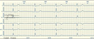
Upon admission, the patient's ECG showed supraventricular tachycardia with a heart rate of 180 beats. per minute Hemodynamics were stable. After examination and examination, the diagnosis was made: “Impaired heart rhythm: Paroxysmal form of left atrial tachycardia with orthograde blockade with a conduction ratio of 1:1, 3:1.” According to Echo-CS, no organic pathology of the heart was detected.
On 12/30/11, due to the ineffectiveness of drug therapy, radiofrequency ablation surgery was performed. After surgical treatment, sinus rhythm was restored.
On January 3, 2012, the patient had a relapse of tachycardia, the heart rate was in the range of 120—140—180 beats per minute. Then the patient was under observation in the cardiology department and was regularly examined by obstetricians and gynecologists. Attacks of tachycardia with a frequency of 180 per minute were accompanied by shortness of breath and were stopped by infusions of a potassium-magnesium mixture.
At 37-38 weeks of pregnancy, the patient was transferred to the department of pathology of pregnant women for delivery by cesarean section. On the second day after delivery, sinus rhythm was restored, and no recurrence of tachycardia paroxysms was observed.
Conduction disorders (slowdown or complete cessation of the conduction of excitation from the sinus node through the conduction system of the heart) during pregnancy usually do not pose a danger. Even with complete atrioventricular block, especially if it is caused by a congenital or acquired heart defect in childhood, pregnancy and childbirth can proceed normally. A contraindication to pregnancy should be considered complete atrioventricular block with a ventricular contraction rate of less than 40 per minute, as well as the presence of Morgagni-Adams-Stokes syndrome.
A particular problem is solving the issue of the possibility of pregnancy and childbirth in women with an implanted artificial pacemaker (APM). IVR with a constant pacing rate does not allow the heart to flexibly respond to changing hemodynamic conditions during pregnancy, which in some conditions complicates the situation. Implantation of an IVR with an adjustable frequency of stimulation allows you to maintain pregnancy, subject to frequent monitoring of the operation of the IVR.
LITERATURE
1. Oganov R. G., Mamedov M. N. National clinical guidelines for cardiology. - M., 2009. - 392 p.
2. Akselrod A. S., Chomakhidze P. Sh., Syrkin A. L. Holter monitoring of ECG. Opportunities, difficulties, mistakes / ed. A. L. Syrkina. - M.: Medical Information Agency LLC, 2007. - 192 p.
3. Tak T., Berkseth L., Malzer R. A case of supraventricular tachycardia associated with Wolff-Parkinson-White syndrome and pregnancy. —Wisconsin Medical Journal. - 2012. - V. 111 (5). - P. 228-232.