Symptoms of myocardial infarction (fever)
The patient’s body temperature on the 1st day of MI usually remains normal and increases on the 2nd, less often on the 3rd day. The temperature rises to 37 - 38 °C and stays at this level for 3 - 7 days. In some cases of extensive heart damage, the duration of the temperature reaction can increase to 10 days. A longer subfebrile condition indicates the addition of complications.
High temperature (39 °C or more) is rare and usually occurs when a complication is associated, such as pneumonia. In some cases, the temperature rises slowly, reaching a maximum after a few days, then gradually decreases and returns to normal. Less often, it immediately reaches its maximum value and then gradually decreases to normal.
The magnitude of the temperature increase and the duration of the fever depend to some extent on the extent of the MI, but the body’s reactivity also plays a significant role in this. In young people, the temperature reaction is more pronounced. In elderly and senile people, especially with small focal MI, it may be insignificant or absent. In patients with MI complicated by cardiogenic shock, body temperature remains normal or even decreased.
The appearance of a temperature reaction after an anginal attack is an important diagnostic sign of MI and should always alert the doctor regarding the development of fresh focal changes in the myocardium. MI is characterized by an increase in the number of leukocytes in the blood. It is observed within a few hours after the development of MI and persists for 3–7 days.
Longer leukocytosis indicates the presence of complications. Usually there is a moderate increase in the number of leukocytes in the blood - up to (10 - 12) * 10 9 /l. Very high leukocytosis (over 20*10 9 /l) is considered an unfavorable prognostic sign.
According to some authors, the severity of leukocytosis to a certain extent depends on the extent of myocardial damage. In approximately 10% of cases, leukocytosis may be normal throughout the entire period of the disease. The number of leukocytes in the blood increases mainly due to neutrophils, and a shift in the leukocyte formula to the left is noted. The first days of the disease are characterized by a decrease in the number of eosinophils in the blood, sometimes even to aneosinophilia. Subsequently, their number increases and returns to normal, and in some cases even exceeds normal levels.
“Myocardial infarction”, M.Ya.Ruda
Read further:
Symptoms of myocardial infarction (presystolic rhythm)
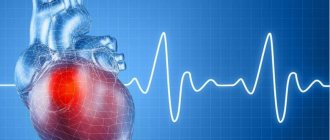
Myocardial infarction
Myocardial infarction is one of the clinical forms of coronary artery disease, which is accompanied by the development of ischemic necrosis of the myocardium due to circulatory disorders in this area. According to statistics, this disease most often develops in males (half as often in females) in the age range from forty to sixty years. The risk of death from myocardial infarction is especially high during the first two hours from its onset
Myocardial infarction - causes
In most cases, myocardial infarction affects people who lead an insufficiently active lifestyle due to psycho-emotional overload. However, a sedentary lifestyle is not a determining factor in the development of this disease, and a heart attack can unexpectedly strike even young people with good physical fitness. The main reasons contributing to the development of myocardial infarction include: bad habits (smoking, drinking alcohol), hypertension, insufficient physical activity, excess animal fats in the food consumed, unhealthy diet, overeating, obesity. Physically active people are several times less likely to develop a heart attack than those who lead a sedentary lifestyle for certain reasons.
The heart is a muscular sac that works like a pump and drives blood through itself. The heart muscle itself is supplied with oxygen through blood vessels that approach it from the outside. For some reason, some of these vessels become clogged with atherosclerotic plaques, as a result of which they cannot pass the required volumes of blood. Coronary heart disease (CHD) develops. Myocardial infarction develops as a result of a sudden complete stop of blood supply to part of the heart muscle due to blockage of the coronary artery. Most often, this is caused by a developed blood clot on an atherosclerotic plaque, much less often by a spasm of the coronary artery. The area of the heart muscle deprived of nutrition dies. Heart attack - dead tissue (lat.)
Myocardial infarction - symptoms
The main typical symptom of this disease is intense pain in the heart and behind the sternum. The pain occurs unexpectedly, in the shortest possible time reaching great severity and “radiating” to the interscapular space, the left shoulder blade, the lower jaw and the left arm. Unlike the pain observed with angina pectoris, pain during myocardial infarction is much more intense and does not go away after taking nitroglycerin (sometimes it is not eliminated even by injections of morphine). In such patients, it is necessary to take into account the presence of coronary artery disease during the course of the disease, as well as the displacement of pain in the left arm, lower jaw and neck. In addition, in older people, this disease can manifest itself in the form of shortness of breath and loss of consciousness.
In 50% of patients, harbingers of a heart attack are angina attacks that change in intensity and frequency. They become more persistent, occur much more often even with slight physical stress (sometimes they can occur even at rest), last longer, and in their intervals in the area of the heart there remains a feeling of pressure or dull pain. Sometimes a heart attack may be preceded not by pain, but by dizziness and general weakness.
In 15% of patients, an attack of pain lasts no more than one hour, in 40% of patients from two to twelve hours, in 45% of patients - about one day.
In some patients, myocardial infarction is accompanied by sudden shock and collapse. The patient turns pale, feels dizzy and severely weak, breaks out in sweat, and may experience short-term loss of consciousness, nausea, vomiting, and diarrhea (rarely). The patient is haunted by a feeling of intense thirst. The skin becomes moist, gradually taking on an ash-gray tint, and the tip of the nose and limbs are cold, blood pressure drops sharply (sometimes it may not be detected at all). The pulse on the radial artery cannot be felt at all or is very weak. During collapse, the number of heartbeats may be slightly reduced, slightly increased or normal (tachycardia is more often observed), body temperature is slightly increased. If collapse and shock continue for many hours and even days, the prognosis for a normal outcome worsens significantly. With myocardial infarction, serious disorders of the gastrointestinal tract can be observed - intestinal paresis, pain in the epigastric region, nausea and vomiting. No less serious disorders can be observed from the central nervous system - fainting, short-term loss of consciousness, general weakness, difficult to eliminate persistent hiccups. With myocardial infarction, serious cerebral circulation disorders may develop, manifested by paresis, convulsions, coma, and speech impairment.
In addition to the specific symptoms described above, patients may experience general symptoms: the number of red blood cells in the blood increases and other biochemical changes are observed, fever appears, body temperature does not exceed the threshold of 38 * C
Clinical forms of myocardial infarction:
— Asthmatic form (the disease begins with an attack of cardiac asthma)
— Anginal form (a heart attack begins with pain attacks in the heart and behind the sternum)
— Abdominal form (begins with dyspeptic symptoms and pain in the upper abdomen)
— Collaptoid form (the disease is preceded by collapse)
— Cerebral form (the disease begins with focal neurological symptoms)
— Mixed form
— Painless form (latent onset of myocardial infarction)
Atypical forms of myocardial infarction
In addition to the tearing, sharp pain behind the sternum characteristic of a heart attack, there are several forms of heart attack that do not manifest themselves at all, or are disguised as other various diseases of the internal organs.
Painless form of heart attack. This form is manifested by a feeling of discomfort in the chest, severe sweating, deterioration of mood and sleep. This form of heart attack most often occurs in old and elderly people, especially with concomitant diabetes mellitus.
Asthmatic form of myocardial infarction. This type of heart attack is very similar in its manifestations to an attack of bronchial asthma and is manifested by a feeling of congestion in the chest and a dry hacking cough.
Gastric form of myocardial infarction. Its symptoms are very similar to an exacerbation of gastritis and are characterized by severe pain in the epigastric region. On palpation, tension and soreness of the muscles of the anterior abdominal wall are noted. In the gastric variant, the lower parts of the myocardium of the left ventricle adjacent to the diaphragm are most often affected.
Diagnosis of myocardial infarction
The diagnosis is established based on a clinical assessment of the patient’s general condition and after differential diagnosis with diseases such as acute pericarditis, dissecting aortic aneurysm, pulmonary embolism and spontaneous pneumothorax. One of the leading diagnostic methods is an electrocardiographic study (ECG), based on the data of which one can judge the localization and extent of myocardial damage and how long ago the process has developed. A heart attack is characterized by changes in laboratory blood parameters: the level of cardiac-specific markers – cardiomyocytes – increases
Myocardial infarction - treatment
The main goal of treating a patient with acute myocardial infarction is to restore and further maintain blood circulation to the affected area of the heart muscle as quickly as possible. The following medications are used for this:
— Acetylsalicylic acid (aspirin) – due to platelet inhibition, the formation of blood clots is prevented
- Prasugrel, Ticlopidine, Clopidogrel (Plavix) - also prevent blood clot formation, but are much more powerful than aspirin
- Bivalirudin, Fraxiparin, Lovenox, Heparin - anticoagulants that prevent the formation and spread of blood clots and affect blood clotting
— Reteplase, Alteplase, Streptokinase are powerful thrombolytic drugs that can dissolve an already formed blood clot
All of the above drugs are used in combination and are vital for the successful treatment of myocardial infarction.
The best modern method of restoring blood flow in the coronary artery is immediate angioplasty of the coronary artery followed by installation of a coronary stent. If for some reason angioplasty cannot be performed during the first hour of a heart attack, the use of thrombolytic drugs is preferable.
If all of the above measures are impossible or do not help, the only means to restore blood circulation (save the myocardium) is urgent coronary artery bypass grafting.
The most critical are the first days of the disease. The further prognosis directly depends on the degree of damage to the heart muscle, the timeliness of measures taken and the presence of concomitant cardiovascular diseases.
More articles on this topic:
Atypical symptoms of cardiovascular pathology
Every practicing physician, regardless of specialty, regularly encounters complex cases of diseases that occur with atypical symptoms. Uncharacteristic manifestations often cause late diagnosis and untimely initiation of adequate therapy, which significantly worsens the prognosis and increases the frequency of deaths among such patients. Pathology of the cardiovascular system most often occurs with various symptoms typical of other localizations of disorders. The reason for this is that the heart muscle and blood vessels provide blood flow with essential nutrients and oxygen to all organs. Insufficient blood supply is naturally accompanied by disturbances in the functioning of one or another tissue, which is manifested by symptoms of the corresponding localization.
Patients do not always feel the well-known attacks of intense chest pain typical of coronary artery disease [1]. The localization may be different: in other parts of the chest, in the area of the shoulder joints, in the abdomen. The close proximity of the heart muscle to the organs located in the epigastrium causes pain in the upper abdomen, which can be observed when the ventricles of the heart expand due to myocardial ischemia, cardiosclerosis, certain heart rhythm disturbances, defects or carditis [2, 3]. A decrease in the left ventricular ejection fraction, regardless of the immediate cause, causes insufficient blood supply to all abdominal organs, which forms the corresponding pathology: ischemic enterocolitis, gastritis, pancreatitis, hepatitis [4]. Dysfunction of these organs is, to one degree or another, accompanied by nausea, sometimes vomiting, changes in stool, and pain, which often becomes a reason to consult a gastroenterologist. The results of endoscopic, ultrasound and laboratory examinations reveal deviations in accordance with one or another pathology. However, standard therapy, in the absence of adequate treatment of cardiac disease, does not give the expected result.
To illustrate the course of ischemic gastroenterocolitis against the background of coronary heart disease, I give clinical example No. 1.
Man Ya., 46 years old, consulted a therapist with complaints of colicky pain throughout the abdomen with an imperative urge to defecate when jogging, which required stopping the exercise. Within a few hours after the workout, I noticed loose, pasty stools 2-3 times over the course of several hours.
From the anamnesis: the pain first appeared about a week ago. During this period, he noted an increase in fatigue, but denied shortness of breath and palpitations. Previously, he was involved in active sports at the amateur level regularly, for 25 years, but did not participate in competitions. Annual planned examinations as part of medical examination were carried out regularly, no chronic pathology was previously identified, and no drugs were prescribed for continuous use.
On examination: condition is satisfactory. The skin is a little pale. There is no swelling. On auscultation, breathing is vesicular, there are no wheezes, the respiratory rate is 17 per minute. Heart sounds are slightly muffled, 78 beats per minute, rhythmic, no murmurs are heard. Blood pressure - 125/80 mm Hg. Art. The abdomen is soft, the liver is not enlarged. Preliminary diagnosis: irritable bowel syndrome. An additional examination was prescribed: clinical and biochemical blood tests; stool tests: coprogram, bacteriological and dysbacteriosis; ultrasound examination of the abdominal organs, esophagogastroduodenoscopy (EGDS), colonoscopy. Therapy: loperamide 2 mg - 2 capsules 2 times a day, probiotics, sedative herbal medicine.
According to the results of an additional examination: there are no deviations from the norm in the clinical blood test. Biochemical blood test: total cholesterol - 5.6 mmol/l, low-density lipoproteins - 4.3 mmol/l, triglycerides - 3.2 mmol/l, aspartate aminotransferase - 56 U/l, alanine aminotransferase - 21 U/l, total bilirubin - 13 µmol/l. Stool tests revealed remains of undigested food, a moderate amount of digestible fiber, otherwise no pathology, no pathogenic agents or dysbiotic changes. An ultrasound examination of the abdominal organs also revealed no pathological changes. According to the results of endoscopy: chronic gastritis, pathological conclusion: chronic moderately severe inactive gastritis without atrophy and metaplasia, test for Helicobacter - negative. According to the results of colonoscopy: no pathology was detected.
After 7 days, despite regularly taking prescribed medications, the complaints persist.
Due to the lack of effect of treatment, significant data for a gastroenterological disease, but at the same time an increase in aspartate aminotransferase with normal values of alanine aminotransferase and total bilirubin [5, 6], an electrocardiographic study (ECG) was prescribed.
ECG: sinus rhythm, 74 beats per minute, normal position of the electrical axis of the heart, supraventricular and ventricular extrasystole. To clarify the nature of the rhythm disturbance, Holter ECG monitoring was performed.
Analysis of the recording revealed frequent supraventricular and ventricular extrasystole of type 4b/5 according to Lown-Ryan, mainly during physical activity, and episodes of sinus tachycardia. 7728 supraventricular extrasystoles were registered, including 25 couplets, as well as 5458 polymorphic (type 3) ventricular extrasystoles, including 12 couplets, 4 triplets.
In Fig. Figure 1 shows a recording section of 24-hour ECG monitoring, containing supraventricular and ventricular extrasystoles in isolated form, as well as a triplet. An example of identified ventricular couplets is presented in Fig. 2, and supraventricular - in Fig. 3. In the ECG recording area in standard lead II for 1.5 minutes (Fig. 4), frequent extrasystole recorded in this patient is visible.
According to echocardiography (carried out against the background of frequent extrasystole): ejection fraction 52% (according to Teicholtz), hypertrophy of the interventricular septum (18 mm), impaired diastolic function of the left ventricle type I, zones of local hypokinesia and akinesia were not identified, there is no evidence of heart disease.
An additional study in the blood serum: MB-creatine kinase - 18 U/l, troponin I - negative, C-reactive protein - 7 mg/l, anti-myofibrillar antibodies to the myocardium were detected in a titer of 1:80, which indicates an inflammatory process in the myocardium.
Based on the data obtained, a diagnosis was formulated: probable myocarditis, supraventricular and ventricular extrasystole. The diagnosis was confirmed by the results of magnetic resonance imaging of the heart with contrast and ECG synchronization, which meets modern requirements for the diagnosis of these diseases [7].
Thus, the presented example demonstrates the possibility of cardiac pathology with symptoms more characteristic of a gastroenterological disease. However, the appearance or increase in complaints with increasing physical activity indicates heart failure as the immediate cause. A decrease in blood supply to the organs of the gastrointestinal tract can occur with a decrease in the ejection fraction of the left ventricle, both due to the inflammatory process in the heart muscle itself, and independently of this, due to frequent ventricular extrasystole. In other cases, this is possible with increasing coronary insufficiency in patients with coronary heart disease. For the purpose of differential diagnosis, it is necessary to pay attention to the condition of the skin, possible swelling, and during auscultation - to a decrease in the sonority of heart sounds, new noises or rhythm disturbances [8]. This will undoubtedly reduce the likelihood of misdiagnosis.
An atypical picture of cardiac pathology can develop with a predominance of completely different symptoms. Impaired blood supply to the brain against the background of various heart pathologies is manifested by typical complaints of dizziness for discirculatory encephalopathy, sometimes fainting, unsteadiness when walking, deterioration in information perception and memorization, noise or ringing in the ears [9]. Possible impairment of vision and/or hearing. In some cases: with endocarditis with the formation of vegetations on the valves and the inner wall of the left ventricle, with atrial fibrillation, severe dilatation of the heart chambers with the absence of adequate antithrombotic therapy, the deterioration of the patient’s condition is associated with thromboembolism. In such situations, the clinical picture usually corresponds to an acute disruption of cerebral blood supply, as confirmed by the results of computed tomography or magnetic resonance imaging. Just as with gastroenterological manifestations, in order to relieve neurological symptoms it is necessary, first of all, to use adequate treatment regimens for cardiac pathology.
A decrease in blood supply to the kidneys naturally leads to the formation of renal failure, which is accompanied by characteristic changes, primarily in laboratory parameters: an increase in creatinine and urea in the blood serum. Other abnormalities are often associated: hyperkalemia, hyperuricemia, hypercalcemia. Complaints of weakness, skin itching, swelling with a predominant localization in the face and with maximum severity after sleep, usually appear later. Signs of renal failure in patients with cardiac pathology, accompanied by a decrease in ejection fraction, are possible in the absence of any independent disease of the renal parenchyma or the collecting system. Successful correction of cardiac parameters in such patients leads to a significant improvement in renal function and relief of clinical symptoms.
Practice shows that patients with cardiac pathology often complain of pain in the joints, back, and muscles. Normalization of the left ventricular ejection fraction and relief of rheological disorders in many cases leads to a noticeable improvement in the subjective and objective characteristics of motor function and a marked decrease in the intensity of pain.
Left ventricular heart failure in any heart pathology is naturally accompanied by an accumulation of blood volume in the pulmonary circulation. Patients are usually bothered by a dry or wet cough, sometimes streaked with blood [1, 10]. It is well known that most often this is manifested by moist, silent rales that are auscultated over the lower parts of the lungs. However, there may be other variants of symptoms, which significantly complicates differential diagnosis. In patients who are in a horizontal position most of the time, wheezing quite often spreads to all parts of the chest, which must be differentiated from developing pulmonary edema. The appearance of fluid in the alveoli gives crepitus, and accumulation in the tissues can lead to compression with narrowing of the lumen of small caliber bronchi, which is accompanied by obstructive wheezing. This situation turns out to be the most difficult, since all the symptoms characteristic of broncho-obstructive syndrome develop: expiratory shortness of breath with wheezing exhalation and reliable signs of obstruction based on the results of an assessment of external respiratory function. A distinctive feature that should be paid special attention to is the insignificant or incomplete effect of the bronchodilators commonly used in such situations. In some cases, due to tachycardia and natural myocardial ischemia developing against their background, health deteriorates with increasing shortness of breath or pain.
To illustrate another variant of the atypical, but at the same time natural course of cardiac pathology, I give clinical example No. 2.
Man K., 39 years old, consulted a therapist with complaints of paroxysmal dry cough, mixed shortness of breath when walking at a moderate pace, and dizziness.
From the anamnesis: deterioration of health was noted within 3 weeks, when coughing and fatigue when walking appeared. I took herbal expectorants on my own, but without effect. Previously, similar symptoms were of concern in acute respiratory diseases. However, unlike previous episodes, this time there was no runny nose, sore throat, or fever. Denies smoking and other bad habits. There were no allergic reactions. No chronic pathology was previously detected.
Upon examination, the condition is closer to moderate. Body temperature - 36.7 °C. Overweight (122 kg with a height of 182 cm) Moderate pasty legs. The skin is slightly pale, the lips are cyanotic. On auscultation, breathing is harsh, carried out in all parts, scattered wheezing in a small amount, in the lower parts - moist, silent wheezing on both sides. The respiratory rate is 22 per minute. Heart sounds are muffled, arrhythmic, heart rate is 106 beats per minute. Blood pressure - 128/92 mm Hg. Art. The abdomen is soft, the liver is +2 cm from the edge of the costal arch.
Preliminary diagnosis: acute obstructive bronchitis.
An additional examination and therapy were prescribed: azithromycin 500 mg per day, probiotics, inhalations with Berodual 4 times a day, Lazolvan 90 mg per day.
Based on the data obtained, it was determined: in the clinical blood test - without pathological abnormalities; in biochemical - total cholesterol - 6.8 mmol/l, low-density lipoproteins - 4.9 mmol/l, triglycerides - 4.2 mmol/l, creatinine - 145 µmol/l, urea - 12.3 µmol/l. The x-ray shows an increase in the vascular pattern, signs of congestion in the pulmonary circulation, a pronounced enlargement of the heart in diameter: cardiothoracic index = 72%.
During treatment, after 5 days the patient notices a deterioration in health, and therefore further examination is carried out to determine the condition of the heart.
The ECG recorded an abnormal supraventricular rhythm with a frequency of 102–140 per minute, atrial fibrillation, repolarization disturbance (ST depression up to 1.5 mm) in the lateral regions.
According to the results of echocardiography, the left ventricular ejection fraction (according to Teicholz) is 44%, pronounced atrial dilatation, moderate ventricular dilatation. There is no evidence of heart disease.
Taking into account the identified changes, a diagnosis was made: atrial fibrillation of unknown duration, cardiomyopathy (?) of unknown etiology, circulatory failure stage II B according to Strazhesko-Vasilenko, functional class III according to NYHA [1, 10]. Rhythm restoration is not indicated due to atrial dilatation.
Treatment was prescribed in accordance with the data obtained: rivaroxaban, carvedilol, starting with small doses; spironolactone, torsemide, maintenance dose of digoxin with heart rate adjustment. With this therapy, the patient's well-being improved significantly. Cough and dizziness do not bother me. He tolerates household physical activity much better.
When examined after 3 weeks: the condition is satisfactory. There is no swelling. The skin is of normal color. Auscultation: vesicular breathing, no wheezing. The respiratory rate is 16 per minute. Heart sounds are muffled, arrhythmic, heart rate is 70 beats per minute. Blood pressure - 118/82 mm Hg. Art. Liver +1 cm from the edge of the costal arch.
During further examination, cardiac magnetic resonance imaging with contrast was performed, which confirmed the presence of dilated cardiomyopathy in the patient.
The presented example shows the possibility of symptoms atypical for cardiac pathology that naturally accompany severe heart pathology in the absence of any significant other disease.
Thus, cardiac diseases can naturally occur with symptoms of pathology of other organs and systems. The correct diagnosis can be made with a thorough analysis of the history, examination and the results of additional examination.
Literature
- Belenkov Yu. N., Oganov R. G. Cardiology: national guide. M.: GEOTAR-Media, 2007. 1232 p.
- Ruzhentsova T. A. Diagnosis and treatment of myocardial lesions in children with acute intestinal infections (clinical experimental study). Author's abstract. dis. ... doc. honey. Sci. M., 2021. 48 p.
- Ruzhentsova T. A. Infectious myocarditis // Practicing doctor today. 2013; 1:48–56.
- Maev I.V., Kazyulin A.N., Kucheryavyi Yu.A. Chronic pancreatitis. M.: Medicine, 2005. 504 p.
- Rosly I. M., Abramov S. V. Principles for assessing enzymological blood parameters in infectious pathology (I message) // Infectious diseases. 2003; 1(1):58–63.
- Rosly I. M., Abramov S. V., Belova E. G., Eremushkina Ya. M. Principles for assessing enzymological blood parameters in patients with infectious pathology (II message): intoxication syndrome // Infectious diseases. 2004; 2 (1): 12–18.
- Caforio AL, Pankuweit S, Arbustini E et al. Current state of knowledge on aetiology, diagnosis, management and therapy of myocarditis. A position statement of the European Society of Cardiology Working Group on Myocardial and Pericardial Diseases // Eur. Heart J. 2013; 34:2636–2648.
- Ruzhentsova T. A. Gastroenterological masks of cardiovascular pathology // Experimental and clinical gastroenterology. 2016; 130(6):114–117.
- Tobias JD, Deshpande JK, Johns JA, Nichols DG Inflammotary heart disease/Ed. DG Nichols et al. Critical Heart Disease in Infant and Children. 2nd ed. — Philadelphia, PA: Mosby. 2006: 899–925.
- Yancy CW, Jessup M, Bozkurt B et al. ACCF/AHA guideline of management of heart failure: a report of the American College of Cardiology Foundation: American Heart Association Task Force on Practice Guidelines // J. Am. Coll. Cardiol. 2013; 62:1495–1539.
T. A. Ruzhentsova, Doctor of Medical Sciences
Federal Budgetary Institution Central Research Institute of Epidemiology of Rospotrebnadzor, Moscow
Contact Information