Resuscitation is a set of measures aimed at restoring the vital functions of the human body. Translated from Latin, “reanimation” means “re-giving life.”
A set of resuscitation actions can be performed by both doctors and ordinary people who find themselves next to a person who is in a state of clinical death. Often the term “resuscitation” is applied to the department where doctors carry out intensive care measures. This term also includes a specialized emergency team that responds to calls to people who are in a state of clinical death.
Contraindications to resuscitation measures
The resuscitation procedure has several contraindications:
- severe illness and critical condition of the body, in which resuscitation actions do not make sense;
- the presence of cadaveric spots on the body;
- the presence of rigor mortis;
- changes in the color of the iris, clouding of the cornea, cat's eye syndrome.
Severe and non-life-threatening injuries are also a reason to refuse resuscitation measures. These include, for example, the tearing off of a large part of the torso with massive bleeding, and the tearing off of the head.
Resuscitation of adults, children and newborns
Cardiopulmonary resuscitation in adult patients is carried out according to a single algorithm, regardless of gender. Its main task is to restore airway patency and independent breathing. Resuscitation efforts may be difficult due to the patient's large body weight. The ratio of the frequency of artificial respiration in relation to chest compressions should be 2:30. The optimal frequency of chest compressions during resuscitation is 80 times per minute.
Pediatric resuscitation is carried out by pediatricians and neonatologists. Before performing it, it is important to check for foreign objects in the mouth by examining the visible part of the pharynx. It is better for a child to perform artificial respiration “mouth to nose”, alternating it with indirect cardiac massage. For children under 10 years of age, the frequency of chest compressions should reach 10 times per minute. Pressing is performed with one hand. The amplitude of chest oscillations should not exceed 3-4 cm.
Restoring the vital functions of the body of newborn babies is the responsibility of neonatologists and pediatric nurses. The complex of resuscitation actions of these specialists has been worked out to the point of automation and is aimed at restoring full blood circulation in the baby.
Criteria for assessing the effectiveness of resuscitation measures
The effectiveness of resuscitation measures is assessed based on the following criteria:
- restoration of the reaction of the pupils to light, their narrowing;
- improvement of skin color;
- reduction or complete disappearance of cyanosis of nails and lips;
- presence of respiratory movements;
- the presence of a pulse in the area of the carotid artery;
- the ability to listen to the heartbeat through the chest.
The patient may remain unconscious even after resuscitation is completed. The main criteria for the life of his body are the resumption of free breathing and heart function. In some cases, the heart rhythm is restored, but breathing is not. Then artificial respiration is required until the emergency team arrives.
Intensive care unit patients
The following categories of patients are admitted to the intensive care unit:
- after surgical operations;
- those who were treated in a specialized department after their condition worsened;
- those who experienced clinical death before hospitalization;
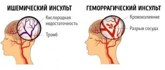
- patients in serious condition, on the verge between life and death, in a coma, after severe poisoning, massive bleeding and injuries, in a state of shock, after a stroke or myocardial infarction.
After stabilizing the condition of patients from intensive care, they are usually sent to specialized departments (therapy, gynecology, surgery, oncology).
Terminal states
With the help of intensive therapy and the use of resuscitation measures, in many cases it is possible to prevent and eliminate the energy and structural deficit that develops in the body during terminal conditions, and save it from death.
Intensive therapy is a set of methods for temporary artificial replacement of vital body functions, aimed at preventing the depletion of adaptive mechanisms and the occurrence of a terminal condition.
Reanimatology is the science of reviving the body; prevention and treatment of terminal conditions (according to V.A. Negovsky).
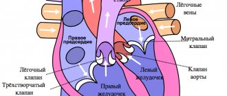
For human life to exist, it is necessary that oxygen continuously enters and is consumed by cells and carbon dioxide is released from it. These processes ensure the coordinated functioning of the respiratory and circulatory organs under the control of the central nervous system. Therefore, their defeat leads to death (“the gates of death” - as defined by the ancients - the lungs, heart and brain).
Stopping vital activity (death) can occur suddenly (in accidents) or predictably, as a natural consequence of old age or an incurable disease. During a long process of dying, the following stages are noted.
Peredagonia . The physiological mechanisms of the body’s vital functions are in a state of deep exhaustion:
- the central nervous system is depressed, possibly comatose;
- heart activity is weakened, the pulse is thready, systolic blood pressure is reduced to a more critical level (70 mm Hg);
- external breathing is weakened, ineffective: tidal volume and frequency are inadequate;
- the functions of parenchymal organs are impaired.
Peredagonia can take several minutes, hours, or even days. During this time, the patient's condition worsens even more and ends with a terminal pause. The patient loses consciousness, blood pressure and pulse are not determined; breathing stops, reflexes are absent. The terminal pause lasts up to a minute.
The next stage of dying is agony (struggle). Due to the depletion of higher-order vital activity centers (CNS), the bulbar centers and reticular formation go out of control (activate). The patient's muscle tone and reflexes are restored, external breathing appears (random, with the participation of auxiliary muscles). The pulse is palpated above the main arteries, vascular tone can be restored - systolic blood pressure increases to 50 - 70 mm Hg. However, at this time, metabolic disorders in the body's cells become irreversible. The last reserves of energy accumulated in high-energy connections quickly burn out and after 20 - 40 seconds clinical death occurs.
In a number of pathological situations (drowning, electric shock and lightning, being hit by a car, strangulation asphyxia, myocardial infarction, etc.), clinical death can befall the victim unexpectedly, without preliminary manifestations of dying.
The main signs of clinical death:
- absence of pulsation over the main arteries (carotid and femoral);
- persistent pupil dilation with absence of photoreaction;
- lack of spontaneous breathing.
Auxiliary signs:
- change in skin color (dead gray or bluish);
- lack of consciousness;
- lack of reflexes and loss of muscle tone.
An important factor influencing the effectiveness of resuscitation in case of clinical death is the ambient temperature and the duration of dying. In case of sudden cardiac arrest, clinical death in conditions of normothermia lasts up to 5 minutes, at sub-zero temperatures - up to 10 minutes or more. A long period of dying significantly worsens the effectiveness of resuscitation, reducing the period of clinical death. Biological death occurs when, as a result of irreversible changes in the body, and above all, in the central nervous system, a return to life is impossible.
Stages and stages of resuscitation
A set of emergency measures carried out to patients in a state of clinical death, aimed at restoring the body’s vital functions and preventing irreversible damage to its organs and systems, is called resuscitation. The person who revives the victim is called a resuscitator.
Returning a patient to a full life from a state of clinical death is possible only with a qualified and consistent implementation of a complex of resuscitation measures.
The first stage of resuscitation is the provision of first aid (basic life support) carried out by a resuscitator (not necessarily a medical worker, but every trained person who has basic resuscitation skills). After clinical death is declared (which should take no more than 7-8 seconds!), preparatory measures are immediately carried out: the victim is laid on his back, preferably with the upper part of his body lowered, on a solid base. A savior who is not involved in intensive care lifts the victim’s legs 50–60 cm upward to drain blood from them and increase blood supply to the cavities of the heart.
The first stage of resuscitation is to ensure airway patency. The resuscitator performs a “triple technique” (according to P. Safar):
a) opens the victim’s mouth and with a finger wrapped in a handkerchief (gauze cloth for clamps) frees him from foreign bodies and liquids (vomit, sputum, algae, inserted crevices, blood clots, etc.);
b) tilts the victim’s head as far back as possible, placing an improvised cushion under the neck (for example, the forearms). In this case, in most victims, the upper respiratory tract is freed from the tongue and its root, becoming open;
c) brings the lower jaw forward. The patency of the upper respiratory tract is restored in other cases.
The second stage of resuscitation is performing mouth-to-mouth artificial ventilation. Having covered the victim's mouth with a bandage (handkerchief), the resuscitator tightly covers his mouth with his lips and exhales. Mandatory condition: the victim's head is tilted back, his nostrils are pinched with the thumb and forefinger (so that the air does not return back), the exhalation volume for adults should be 800 - 1000 ml. When blowing air, the resuscitator, out of the corner of his eye, watches the movements of the victim’s chest, which should rise and fall. Having made 2 - 3 exhalations, the savior begins to carry out the next stage of resuscitation.
The third stage of resuscitation is closed cardiac massage. Being on the side of the victim, the resuscitator places one hand on the lower third of the sternum, strictly in the middle, so that the fingers are raised up and placed parallel to the ribs. He places the second hand on top and, pressing rhythmically, moves the sternum in the sagittal direction to a depth of 3 - 5 cm. The frequency of pressing is 60 per minute.
Mandatory condition: when pressing on the sternum, the fingers of the hand should be raised up to prevent fractures of the ribs, and the arms should be straightened at the elbow joints. Cardiac massage is thus carried out by the mass of the resuscitator’s torso. Subsequently, the rescuer alternately injects air and presses on the sternum in a ratio of 1: 4. If there are two resuscitators, each of them performs its own stage of resuscitation. If necessary, each time remove the lower jaw to ensure patency, breathe in the 2nd and 3rd stages in a ratio of 2: 10.
Signs of effective resuscitation measures :
- constriction of the pupils,
- normalization of skin color,
- feeling under the fingers of arterial pulsation, synchronous with the massage;
- Sometimes blood pressure is determined.
In some cases, cardiac activity may be restored. The first stage of resuscitation should be carried out continuously until the arrival of a specialized medical team.
The second stage of cardiopulmonary resuscitation is the provision of specialized medical care (further life support). It is carried out by professional doctors using monitoring, diagnostic, therapeutic equipment and medications. Under the control of a laryngoscope, a portable or electric suction is used to better clean the airways and intubate the trachea.
The resuscitation team provides more effective artificial ventilation of the lungs - with a manual portable or stationary ventilator through a mask, air duct or endotracheal tube with an air-oxygen mixture. If you have a special massager, you can perform closed cardiac massage using hardware.
At this stage, the following stages of resuscitation are carried out alternately. The first stage is drug therapy. For all types of circulatory arrest, solutions of adrenaline hydrochloride (1 ml of 0.1% solution) and atropine sulfate (3 ml of 0.1% solution) are used. The second stage is assessing the type of circulatory arrest. For adequate drug therapy, it is necessary to diagnose the functional state of the victim’s heart. To do this, the patient is connected to an electrocardiograph (cardioscope) in the second lead and the curve is recorded.
There are the following types of circulatory arrest : asystole, fibrillation, “ineffective heart”.
- During asystole, the electrocardiograph records a straight line.
- Ventricular fibrillation is manifested by frequent chaotic contraction of individual myocardial fibers (uneven “teeth” of high, medium and variable amplitude).
- “Ineffective heart” - registration of the ventricular complex on the ECG in patients with absent pumping function of the heart.
Ventricular fibrillation and an “ineffective heart” without appropriate correction, as the energy resources of the myocardium decrease, quickly turn into asystole. In the presence of high-wave fibrillation, you can use a 2% lidocaine solution (0.5 mg per 1 kg of body weight, repeatedly).
In case of an “ineffective heart” caused by a sharp decrease in the volume of circulating blood, to ensure its pumping function, hemodynamic media (refortan, stabizol, polyglucin, rheopolyglucin), crystalloids, and glucocorticoids are infused intravenously or intraarterially; in case of massive blood loss - canned blood and its components.
In case of cardiac arrest caused by hyperkalemia or hypocalcemia (acute and chronic renal failure, hemolysis of red blood cells, massive tissue destruction, hypoparathyroidism), calcium chloride should be used (5 - 10 ml of 10% solution, intravenously). Medicinal media are administered intravenously, into the lumen of the trachea, through a cannulated artery, intracardiacly.
Medians administered intravenously, with effective cardiac massage, enter the coronary vessels, exerting their influence on the heart. If it is impossible to provide venous access, the intratracheal method is used, which is no less effective. In this case, with a needle for intramuscular injections, the cricothyroid ligament or the space between the tracheal rings is pierced and solutions of adrenaline, atropine or lidocaine are injected (2-3 ml of 0.1% solution of adrenaline hydrochloride, 1 ml of 0.1% solution of atropine sulfate, 2 ml of 2% lidocaine solution - diluting them in 10 ml of sodium chloride solution). These medications can also be given through an endotracheal tube. During ventilation of the lungs at this time, medicinal media penetrate through the alveoli, and after 30-40 seconds enter the lumen of the coronary vessels.
According to some researchers, using distilled water as a solvent can achieve effective absorption of the solution from the alveoli into the blood due to the difference in osmotic pressure. Due to the large number of complications, intracardiac administration of drugs is used less and less (the technique is described below).
The intra-arterial method of infusion in patients with a significant loss of circulating blood volume (for example, with hemorrhagic shock and an “ineffective heart”) also deserves attention Infusion of blood and other agents into a catheterized artery (eg, radial) is often effective in filling the coronary arteries with blood, ensuring efficient cardiac function and restoring hemodynamics.
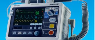
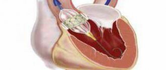
The third stage is electrical defibrillation of the heart. It is used in patients with ventricular fibrillation (the method is described below). In an operating room, it is sometimes more effective to perform open cardiac massage. Indications: cardiac arrest during surgery on the organs of the chest and upper abdomen; cardiac tamponade; bilateral pneumothorax; multiple fenestrated rib fractures; congenital and acquired deformities of the chest, which make it impossible to conduct effective closed massage. Open massage is carried out through transthoracic or subdiaphragmatic access, without opening or with opening of the cardiac membrane.
A prerequisite for this is that the fingers of one or both hands, when squeezing the ventricles, should be placed along the vascular bundle so as not to pinch it. The third stage of cardiopulmonary resuscitation.