Pelger neutrophil anomaly is a benign hereditary pathology that is characterized by morphological changes in leukocytes. It was named after Karl Pelger, a Dutch doctor who first discovered atypical neutrophils in 1928. Then in 1932, Huett proved the hereditary nature of the disease. Therefore, in some sources the disease is called Pelger-Huet anomaly. Occurs with a frequency of 1:1000-1500, equally common in men and women. Previously, it was rare, but recently it has become more common, due to more extensive blood examinations.
Neutrophils: general information
Not every patient knows about such a pathology as Pelger anomaly of neutrophils. What kind of disease is this? To answer this question, let's try to understand the structure and functions of neutrophils.
Neutrophils are the most numerous type of white blood cells. They are necessary for the body to fight pathogens of infectious diseases. These cells rush to the site of inflammation. and then absorb and digest dangerous bacteria and viruses. This process of neutralizing foreign agents is called phagocytosis.
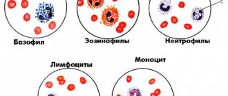
The blood test usually indicates 4 types of neutrophils:
- Myelocytes. These are the youngest cells. Normally, they can only be present in the bone marrow. Their presence in peripheral blood always indicates pathology.
- Young cells. They may be present in a blood test in very small quantities (up to 1% of the total number of neutrophils);
- Band neutrophils. This is a small group of cells. They are 1 - 5% normally.
- Segmented cells. It is this type of neutrophils that predominates in the peripheral blood (40 - 70%).
Mature types of neutrophils include only segmented cells; all other varieties are considered young forms.
With Pelger's anomaly of neutrophils, changes in the structure of segmented cells are noted. They become similar to young forms. This often leads to errors in diagnosing various pathologies.
How does it manifest?
Before studying the manifestations of neutrophilic changes in the blood, you need to understand what function these cells perform. Neutrophils are leukocytes, blood cells that are responsible for ingesting pathogenic bacteria. As soon as pathogenic bacteria or other pathogens enter the blood, neutrophils immediately begin to attack them.
Important! The normal content of segmented neutrophils in the blood is from 45 to 65% in the leukocyte formula. If we talk about the absolute value, the number of neutrophils can vary from 2.0 to 5.5 Giga/liter.
Such an anomaly of leukocytes does not have specific clinical expressions, because their functionality is not impaired. A Pelger anomaly can be detected completely by accident when a person takes a test. Often in people who are carriers of this pathology, ESR and coagulability indicators remain normal. People with this abnormality have the same reactions to blood loss or infection.
Read also: What can cause elevated white blood cells in the blood of newborns and how to normalize their level?
Blood picture
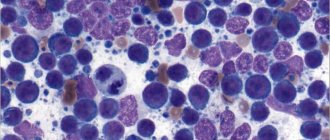
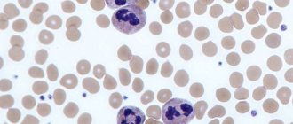
This congenital disorder can only be detected using a clinical blood test. If the nuclei of mature cells have few segments and change shape, this may indicate a Pelger anomaly of neutrophils. In the photo below you can see pathological changes in the nuclei.
The nucleus of Pelger neutrophils can have the following forms:
- oval;
- rod-shaped;
- horseshoe-shaped;
- bean-shaped;
- rounded.
In more rare cases, there are cores with a constriction in the center, similar to a gymnastic weight.
In this case, the core looks too short and consists of only one segment. It has a lumpy structure. Less common are nuclei of altered shape, having two or three segments.
If the nucleus of a segmented neutrophil has a round shape, then it can very easily be mistaken for a myelocyte. Only a detailed blood test can show that this is not a young, but a mature cell. The presence of neutrophils with altered nuclei often leads to errors in the interpretation of analysis results.
Medicine / 7. Clinical medicine
Stoyanova M.M., Volkova S.D.
Institution "Krivoy Rog City Hospital No.
4 "
Pelger's anomaly and pelgeroid
Pelger-Huet anomaly (Pelger familial leukocyte variant) is a benign hereditary condition in which the segmentation of leukocyte nuclei is impaired.
This anomaly was first described in 1928 by the Dutch doctor Pelger, and in 1932 Huet determined its hereditary nature.
Carriers of the anomaly are practically healthy people, since functionally Pelger’s leukocytes are absolutely complete. It occurs in families and is inherited as an autosomal dominant trait. The pathogenesis and etiology of Pelger's anomaly are unknown.
Pelger's anomaly is characterized by two morphological features: hyposegmentation of nuclei and a coarse, coarse, almost pyknotic chromatin structure. Changes are present in leukocytes of all lineages including neutrophils, eosinophils, basophils.
In neutrophils, these changes are most visible and are expressed mainly in changes in the morphology of the nuclei, namely, in the disruption of their segmentation, when a nucleus that is mature in structure has a youthful shape. Most Pelger neutrophils have monolobed, unsegmented nuclei in the form of an ellipse, bean, or short thick rod. There are also bisegmental nuclei in the form of glasses, gymnastic weights, hourglasses with short bridges of different thicknesses. Nuclei with three segments are rare. Along with non-segmented band and segmented neutrophils, round-nuclear neutrophils are also observed, which are recognized as fully mature cells. Some Pelger neutrophils have large, abundant granularity, while in others it is small and scanty.
To avoid erroneous interpretation of the analysis in the presence of the indicated forms of neutrophil granulocytes, the laboratory doctor is obliged to give a conclusion that the described picture is characteristic of Pelger’s leukocyte anomaly. This will allow you to avoid erroneous interpretation of the blood picture “as a left shift” of neutrophils and incorrect tactics of the attending physician. The hereditary nature of changes in leukocyte morphology can be definitively confirmed in a patient after examining the blood of immediate relatives.
Morphologically similar changes not related to hereditary nature (pelgeroids) are described in a number of pathological conditions and diseases as a transient phenomenon.
These conditions include: agranulocytosis, sepsis, malignant tumors, leukemia, radiation sickness, some infectious diseases, malaria, severe forms of intestinal infections. Some authors believe that Pelger cells can appear in the peripheral blood long before the manifestation of hemoblastoses and myelodysplastic syndrome.
In contrast to the true Pelger anomaly, with pelgeroid, the morphological changes in leukocytes are less pronounced, and the structure of the nuclei is less dense. This symptom is defined as pseudopelgerization and also as reactive pelgerization. It differs from the real Pelger anomaly in that these changes in leukocytes disappear after the underlying disease is cured.
In recent years, descriptions of acquired forms of hyposegmentation of the nuclei of neutrophil granulocytes - pelgeroids - have appeared when taking certain medications.
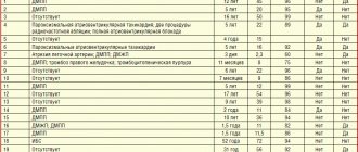
We observed pseudopelgerization of neutrophils in the peripheral blood associated with taking the drug paclitaxel in patient P., 53 years old, diagnosed with carcinoma of the right breast.
After surgical removal of the tumor, the patient took paclitaxel according to the regimen. Over the course of 4 weeks, characteristic morphological changes in neutrophilic granulocytes were repeatedly noted in the leukogram. A large number of short thick rods with a coarse lumpy nucleus structure and bisegmental neutrophils with lumpy nuclei and thin bridges appeared. Cells with three segments were absent. During this period, laboratory tests of the patient’s peripheral blood looked like this: Er - 4.10 - 4.22×10¹²/l, Hb - 125-126 g/l, L - 3.4-4.9×10⁹/l, ESR — 20-25 mm/h. Leukogram: band neutrophils - 18-22%, bisegmented neutrophils - 45-47%, eosinophils 1-2%, lymphocytes 26-30%, monocytes 4-6%. After completion of treatment with paclitaxel, the above-described morphological changes in neutrophils were not observed. Laboratory data are as follows: Er - 4.10 1012/l, Hb - 122 g/l, L - 4.1 109/l, ESR - 10 mm/h, In the leukogram there are band neutrophils - 6%, segmented neutrophils (mainly with three segments) - 54%, eosinophils - 1%, lymphocytes - 34%, monocytes - 5%.
Thus, underestimation of the importance of the morphological characteristics of cells can lead to both overdiagnosis with subsequent unjustified interventions, and to underdiagnosis, which can negatively affect the prognosis of the disease.
Literature.
1. Abramov M.G. Hematological atlas. Moscow “Medicine” 1979 pp. 188-191
2. Vorobyov A.I. (ed.) Guide to hematology. T1 Moscow 1985 With. 415
3. Kamyshnikov V.S.. Methods of clinical laboratory research., 5th edition. Moscow 2011 "MEDpress-inform", p. 266-267
Causes
The anomaly occurs due to a violation of the formation of segments of neutrophil nuclei. The cause is a failure at the genetic level.
This disease is hereditary. It is transmitted in an autosomal dominant manner. This means that the pathology can be passed on to children if both parents are carriers of the defective gene.
The anomaly occurs in 1 child out of 1000 newborns. It is observed equally often in boys and girls. The pathology is benign. Many doctors consider Pelger's anomaly not a disease, but a constitutional feature.
Manifestations
There are no pathological symptoms with Pelger's anomaly of neutrophils. This congenital feature does not affect a person’s well-being in any way and does not pose a health hazard.
Changing the shape of neutrophil nuclei does not affect phagocytosis. Such cells retain the ability to fight foreign microorganisms and produce enzymes. This anomaly does not affect the state of immunity in any way.
A very small proportion of doctors believe that patients with Pelger's neutrophil anomaly are more likely to have short stature and a tendency to have bowed bones. However, these data have not been scientifically confirmed. Most people with this feature are of normal height and do not suffer from bone pathologies.
Clinical manifestations
Clinically, the disease is not expressed in any way, since the functions of leukocytes are not impaired. Leukocytes are also capable of phagocytosis of foreign cells and contain a set of enzymes identical to normal leukocytes. ESR is not increased, blood clotting is normal. Often this pathology is discovered by chance during a blood test. Due to the fact that there is a neutrophil shift to the left in the leukocyte formula, an infection can be mistakenly assumed. Reactions to blood loss, infection, etc. in people with this anomaly the reactions do not differ from healthy people.
The leukocyte formula is the ratio of several types of leukocytes in the human body
According to some sources, there may be changes in the skeletal system: hyperkyphosis, short stature, skeletal deformities.
Consequences
What does Pelger's anomaly of neutrophils lead to? This violation does not cause any complications or negative consequences for human health.
Difficulties arise only when interpreting the results of a blood test. Very often, doctors mistake modified segmented neutrophils for band neutrophils. An increase in con can be a sign of the following diseases:
- oncological diseases of the hematopoietic system;
- bacterial and viral infections;
- injuries and burns;
- myocardial infarction;
- inflammatory processes in various organs;
- allergic reactions;
- sepsis;
- parasitic diseases;
- renal failure.
If the blood picture is altered, the doctor may suspect the above pathologies in the patient. In the history of medicine, there have been cases when patients with Pelger's neutrophil anomaly were mistakenly diagnosed with infectious diseases or leukemia. As a result, people underwent a course of treatment that was absolutely unnecessary for them.
This congenital feature often leads to erroneous interpretation of clinical blood test data. However, a qualified and experienced doctor never makes a diagnosis based on just one test.
Diagnostics
If the patient’s clinical analysis shows signs of Pelger’s anomaly, the doctor must indicate this in the transcript. To avoid errors in diagnosis, a person needs to additionally take a blood smear for the following indicators:
- kernel maturity;
- chromatin density;
- neutrophil ratio.
With Pelger's anomaly, the patient's chromatin density remains normal, the nucleus has an altered shape, but is old. This study helps distinguish altered mature cells from young forms.
If possible, a clinical test and blood smear should be taken from the patient's parents. Since the pathology is hereditary, the same anomaly is observed in the mother and father of the patient. This is an important diagnostic sign.
Is treatment necessary?
Clinical manifestations of Pelger's neutrophil anomaly are completely absent. A person with this feature is considered absolutely healthy and does not need treatment.
It is advisable to identify the anomaly in childhood. This will help avoid diagnostic errors in the future. If a patient with a Pelger neutrophil anomaly is scheduled to undergo a blood test, it is very important to tell the doctor about his congenital abnormality. Otherwise, the test results will be misinterpreted.
Causes and laboratory signs
Unfortunately, the causes of this anomaly have not yet been established, but the mechanism of hereditary transmission is known; it is an autosomal dominant type. It can manifest itself to the same extent in homozygous and heterozygous probands. Its development is based on a special change in the leukocyte nuclei.
It is known that ordinary, mature neutrophils have a segmented nucleus, while pelgerization of neutrophil nuclei occurs due to a violation of the segmentation processes. As a result, the kernels retain their youthful shape upon the onset of full maturity.
If in normal neutrophils the nuclei are divided into several lobules, usually four or five, then with Pelger’s anomaly, if you look under a microscope, then mature and fully functional neutrophils have a nucleus that has only two lobules, rarely three. This lobe has a constriction in the middle, so this core resembles either a peanut, or a gymnastic dumbbell, or pince-nez. In any case, no matter how many segments the nucleus of a Pelger leukocyte contains, it has one more feature. This core has a lumpy structure, and the bridges between the lobes are quite short.
Such abnormal neutrophils are very similar to young leukocytes - myelocytes of normal structure, and this can be a mistake for laboratory doctors, and even for a hematologist. At the same time, whatever the variants of the nuclear structure, Pelger neutrophils are absolutely healthy and full-fledged cells. They can also perform phagocytosis, engulf and digest harmful microorganisms, contain as many enzymes as needed, live as long as normal white blood cells, and respond to all pathological processes in the same way.
Similar anomalies
We examined the congenital type of Pelger anomaly. However, similar changes in neutrophils may also be observed in the following diseases:
- systemic lupus erythematosus;
- tuberculosis;
- malaria;
- flu;
- lymphogranulomatosis;
- decreased thyroid function;
- leukemia
In these cases, the changes are acquired. They are not observed constantly, but only with exacerbation of the underlying pathology.
There is also a very rare congenital disorder that geneticists call SOPH syndrome. It was first described in 2009. This pathology occurs among the peoples of the North, mainly among the Yakuts. Pelger's anomaly is only one of the manifestations of this syndrome. This genetic disease is accompanied by other pathological signs:
- short stature;
- sagging skin;
- atrophy of the optic nerves and retina.
If a child is diagnosed with a Pelger anomaly, then you should take a close look at his well-being and state of health. If no other pathological signs are observed, then there is no cause for concern. This constitutional feature does not affect either the duration or quality of life.