Pathogenesis of vascular disorders in Kimerli anomaly
To understand why Kimerli's anomaly is a serious pathology, it is necessary to first consider the structure of the spine. It is worth knowing that the left and right vertebral arteries arise from the subclavian arteries and then pass along the cervical spine. In this case, these arteries are located in a canal, which is formed by the openings of the vertebral processes. The vertebral artery then enters the foramen magnum and enters the skull.
The vertebral arteries, together with their branches, form the vertebrobasilar system, which supplies the cerebellum, brain stem and spinal cord. These arteries also bend around the cervical vertebra, after which they pass through a wide bony groove. If a patient has a Kimerli anomaly, a bone arch is formed above the bone groove, which limits the movement of the vertebral artery in this place.
Kimerli's anomaly is almost always a congenital pathology. But it does not always provoke serious health problems for the patient. Typically, clinical manifestations of pathology that worsen the patient’s quality of life occur against the background of the following diseases:
- vasculitis;
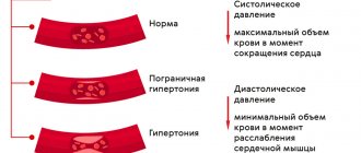
- arterial hypertension;
- atherosclerosis;
- cervical spondyloarthrosis;
- cervical osteochondrosis;
- presence of craniovertebral malformations;
- traumatic brain injury;
- scarring process;
- spinal injury.
When should you see a doctor?
If a person suddenly experiences numbness or tingling in the shoulder, arms or legs, or if the person has lost bowel or bladder control, then seek medical help as soon as possible!
If discomfort and pain begin to interfere with daily activities, it is necessary to consult a neurologist. Despite the fact that spondylosis is part of involutional changes, there are nevertheless various treatment methods that can improve well-being and reduce symptoms.
Classification of Kimerli anomaly
The correctly selected method of treating a disease is largely determined by its type. In neurology, it is customary to distinguish two main types of Kimerli anomaly. The first is characterized by the formation of a bone arch that connects the process of the atlas with the posterior arch. The second type of anomaly involves the presence of a bone arch located between the transverse and articular processes of the atlas.
There are also complete and incomplete types of pathology. With a complete anomaly, the shape of the bone arch resembles a semi-ring, and with an incomplete anomaly, it resembles an arched outgrowth. Kimerli's anomaly can be localized both on both sides of the vertebra and on one side. It is also common to distinguish between acquired and congenital forms of the disease.
As for the acquired pathology of Kimerli, it is usually formed against the background of various chronic pathologies of the spine - for example, with osteochondrosis. In patients with an acquired anomaly, there is usually such a deformation of the groove in which it is closed from above by a ligament.
Kimerli's congenital anomaly is a consequence of developmental defects and does not pose a serious danger to the patient, therefore it does not require special treatment. On the contrary, the acquired form of the disease requires complex treatment, since it is characterized by severe symptoms that threaten the health and life of the patient. The most severe cases of the disease are accompanied by neurological disorders due to poor blood supply and compression of the spinal artery.
What's special?
Kimmerle's anomaly is a pathologically altered structure of the atlas, the first vertebra. Unlike other parts of the spinal column, it does not have a bone base, and looks like a ring of 2 arches. In the posterior arch there is a hollow through which a large blood vessel passes. In case of Kimmerle violation, this groove is blocked by a bone septum, which normally should not exist. Compression of the spinal artery occurs, which disrupts the nutrition of the brain and damages the nerve endings.
According to statistics, such changes in the cervical segment of the spinal column are diagnosed in 12–30% of people, but signs of anomaly are found in only a few. Therefore, this type of atlas is not considered a disease.
Symptoms of Kimerli anomaly
Various manifestations of the disease, which cause severe discomfort to the patient, are usually triggered by a decrease in blood flow to the posterior parts of the brain. As a result of this pathological process, the patient experiences noise in one ear or both. The nature of this noise can be different - hum, whistle, ringing, hissing. For the same reason, there is flickering or flickering before the eyes, as well as sudden darkening in the eyes.
Kimerli's anomaly is characterized by impaired blood supply to the cerebellum, which leads to such clinical manifestations of the disease as frequent dizziness, lack of coordination and unsteadiness of gait. Usually, all these conditions only intensify when turning the head. Constant muscle strain and uncomfortable head position lead to the patient often losing consciousness. Another unpleasant symptom of the disease is the appearance of sudden muscle weakness, which leads to the patient falling without a previous loss of consciousness.
Severe cases of the disease are characterized by more complex and unpleasant symptoms. In patients with severe forms of Kimerli's anomaly, clinical signs of the disease are observed, such as tremor of the arms and legs, headache, nystagmus, hypoesthesia of the face or trunk, coordination disorders, and motor disorders of the extremities. The most life-threatening complication is ischemic stroke.
Clinical picture
Kimmerle's anomaly usually does not appear. With significant compression of the vessel, the nutrition of the brain is disrupted. This is accompanied by the following symptoms:
PAY ATTENTION! Normalize the functioning of your joints and don’t push yourself into a wheelchair, it’s better to be on the safe side, but for this you will need it. normalize >>
- headache;
- decreased visual acuity;
- hearing impairment;
- blood pressure surges;
- manifestations of VSD.
Such a clinical picture causes incorrect diagnosis. The patient is struggling with migraine or vegetative-vascular dystonia. Unsuccessful treatment and worsening of the problem require careful diagnosis to identify the true cause of the ailment. In 20% of people with Kimmerle's anomaly, the disorder is accompanied by symptoms of cerebral ischemia. These include:
Cerebral ischemia is accompanied by “floaters” before the eyes.
Acute disturbances in the blood supply to the brain are accompanied by visual hallucinations and sudden loss of consciousness.
Diagnosis of Kimerli anomaly
Kimerly's anomaly is a serious neurological disease, the effective treatment of which depends on timely and accurate diagnosis. Of great importance in diagnosing pathology is the collection of anamnesis and neurological examination of the patient. Typically, patients consult a doctor with symptoms of Kimerli's anomaly, which indicate a disruption of the circulatory process in the vertebrobasilar region. The doctor primarily prescribes diagnostic methods such as radiography of the skull and spine. These techniques are very effective, since Kimerli's anomaly is quite clearly visible on lateral radiographs of the craniovertebral zone.
If a patient complains of tinnitus, the doctor needs to exclude possible ENT diseases: cochlear neuritis, labyrinthitis, chronic otitis media. For this purpose, it is advisable to refer the patient for examination to an otolaryngologist. Differential diagnosis also involves conducting various hearing tests, such as experiments with tuning forks, speech, noise and tone audiometry. Additionally, studies of the vestibular apparatus may be prescribed: electronystagmography, vestibulometry, stabilography.
Kimerli's anomaly is not always provoked by vertebral artery syndrome, so the neurologist will need to exclude other causes of this pathological process during diagnosis. For example, using contrast angiography, it is possible to detect compression of a vessel by formations such as a cyst, tumor, brain abscess, arteriovenous malformation, thrombosis, and cerebral aneurysm.
Diagnosis of the disease also involves determining the degree of its influence on blood circulation in the vertebrobasilar region. This can be done with the help of additional studies such as transcranial Doppler sonography, ultrasound of extracranial vessels, MRI and duplex scanning of cerebral vessels. All these instrumental techniques make it possible to determine the location of compression of the vertebral artery and disruption of its functioning, depending on the position of the neck and head.
Recovery prognosis
Treatment of a disease does not always bring the desired result. Conservative therapy can improve the patient’s condition and make blood flow more active, but a number of symptoms may still persist . For example, ringing or noise in the ears is difficult to correct, and many patients have to put up with it. As for the prognosis for life, it is usually favorable. In the absence of pronounced symptoms and timely treatment of minimal blood flow disorders, nothing threatens a person’s life.
Treatment of Kimerli anomaly
Treatment of Kimerli's anomaly is not necessary in all cases. Indications for treatment may include serious clinical symptoms of circulatory disorders caused by the disease. Patients in whom Kimerly's anomaly has caused vascular insufficiency are usually prescribed conservative treatment. In particular, to improve cerebral blood flow, vascular therapy is prescribed, which involves taking drugs such as Cavinton, Sermion, cinnarizine, Devincan.
To improve real blood parameters, trental and pentoxifylline are prescribed. Complex therapy for the disease also involves taking neuroprotectors, antioxidants, nootropics and metabolic medications (ginkgo biloba, piracetam, mildronate, picamilon).
Epidemiology
Cervical spondylogenic myelopathy is the most common cause of non-traumatic spastic paraparesis and quadriparesis. Almost 23.6% of patients with non-traumatic myelopathic symptoms had cervical spondylogenic myelopathy.
Cervical spondylosis is more common in men.
The results of studies using radiographic data showed that spondylous changes are most common in people over 40 years of age. After all, more than 70% of men and women suffer from spondylosis in adulthood, but radiographic changes are more pronounced in men than in women.
Surgery
In most cases, treatment of Kimerli's anomaly is limited to conservative methods and does not require surgical intervention. The need for surgery may be due to a decompensated manifestation of vertebral artery syndrome, which provokes a serious circulatory disorder in the vertebrobasilar region. During surgery, the doctor performs mobilization of the vertebral artery and resection of the pathological arch. After the operation, the patient will need to undergo a rehabilitation period, during which he will have to wear a Shants collar for 2-4 weeks.
Risk factors
The biggest risk factor for developing cervical spondylosis is aging. Cervical spondylosis is usually associated with involutional changes in the joints of the neck. Disc herniation, degeneration and bone growths (osteophytes) are all associated with involutional changes in the body.
Other risk factors:
- neck injuries
- heavy lifting work that places additional strain on the neck
- holding the neck in an awkward position for a long time or repeating the same neck movements throughout the day
- genetic determination
- smoking
- overweight and inactivity
Additional measures
Patients diagnosed with a mild form of Kimerly's anomaly should definitely adhere to certain precautions that will help them avoid serious complications. Namely, they need to avoid in every possible way sharp turns of the head, excessive physical exertion, performing physical exercises while standing on their heads, as well as sports games and activities that involve hitting their heads. Such activities include football, gymnastics, basketball and others.
Before a session of manual therapy or massage, the patient must warn the attending physician about the presence of Kimerli's anomaly. He must remember that in the event of a sharp deterioration in his health, he needs to urgently consult a neurologist for advice so as not to miss the early stage of the disease and begin treatment on time.
What's the forecast?
With Kimmerle anomaly, treatment leads to positive results. Often people are unaware of the presence of a violation. If pathology occurs, the risk of ischemia may increase. Symptoms can reduce a person’s quality of life and ability to work, but this rarely happens. The patient is recommended to systematically consult with a neurologist and keep osteochondrosis under control in the case of an acquired form of the anomaly. As prescribed by the doctor, a course of massage is carried out, and an individual system of physical exercises is developed.
In combination with osteochondrosis, many anomalies of the cervical spine have a negative physiological effect on the general condition of the body. As a rule, congenital structural disorders do not appear until concomitant pathology develops. One of these diseases is Kimmerle's anomaly (AK).
se up
Physical examination
First, the doctor finds out the presence of symptoms, their intensity, location, and history of the disease. A neurological examination is then performed, which includes examining reflexes, testing muscle strength, and determining sensory deficits and range of motion in the neck. A doctor may perform a gait analysis to determine how much damage to the spinal cord is present.
If a doctor suspects cervical spondylosis, he will order imaging tests and neurophysiological studies to verify the diagnosis.
Visualization methods
- X-rays can be used to visualize bone spurs and other abnormalities.
- A CT scan can provide more detailed images of the neck.
- MRI scans, which create images using radio waves and a magnetic field, help doctors detect compression of nerve structures.
- Myelogram - This procedure uses contrast to be injected into specific areas of the spine. A CT scan or x-ray is then used to take more detailed images of these areas.
- EMG (ENMG) is used to test the functionality of nerve fibers, the method checks the electrical activity of the nerves and the conduction of impulses to the muscles.
- ENMG determines the speed and strength of the signals that the nerve sends. This allows you to determine the presence and level of damage to nerve fibers.