The Euroonko clinic in Moscow regularly installs vena cava filters for patients with cancer to prevent the development of pulmonary embolism (PE). We also install vena cava filters in patients with a high risk of developing pulmonary embolism who do not suffer from cancer, for example, with deep vein thrombosis of the leg, with various types of blood clotting disorders and a number of other diseases. The information below will be useful for all patients requiring vena cava filters.
There is no single generally accepted concept regarding surgical prevention of pulmonary embolism in international practice. In the USA, a radical approach has been adopted with the installation of vena cava filters, taking into account the embologenicity of the thrombus. In Western European countries, implantation is recommended only if it is impossible to carry out anticoagulant therapy for pulmonary embolism.
Timely placement of vena cava filters in patients with cancer is part of complex therapy for late stages of cancer, and is quite widely used in routine clinical practice in Western Europe. Depending on the form of cancer and the stage of cancer, vena cava filters are installed in approximately 1.5–6.5% of all patients.
Unfortunately, in domestic clinical practice, due to the low financial resources of the healthcare system, the indications for the placement of vena cava filters are significantly narrowed, which contributes to a high mortality rate and a higher incidence of sudden death compared to data from the US Cancer Registry and leading national cancer registries. countries of Western Europe.
Placement of vena cava filters in the inferior vena cava in the early stages of cancer in patients undergoing active chemotherapy, including high-dose chemotherapy, can reduce the risk of sudden death due to the development of thromboembolism, including pulmonary embolism, by approximately 10–15%.
Installation of a vena cava filter allows not only to avoid pulmonary embolism during therapy with a number of chemotherapy drugs, but also to perform thrombolysis of previously occurring blood clots.
The doctors of the Interventional Surgery Department at Euroonko have accumulated extensive experience in the prevention and treatment of venous thrombosis, pulmonary embolism, thrombophlebitis against the background of varicose veins in cancer. The interventional surgery department uses both temporary and permanent venous filters.
What is a vena cava filter?
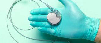
A vena cava filter is a high-tech mesh on a complexly modeled frame or other complex structure. Using X-ray vascular intervention, a vena cava filter is installed into the lumen of the inferior vena cava, just below the entry of the renal arteries into it (infrarenal position).
A vena cava filter installed in the inferior vena cava filters all particles larger than 2–4 mm (depending on design). This allows venous blood flow from the lower extremities and all internal organs to pass freely through the filter, while retaining large clots that could potentially lead to significant thrombosis or PE.
Under the influence of the blood anticoagulation system and anticoagulation drugs taken, for example, warfarin, stuck blood clots dissolve directly on the vena cava filter. If the filter is installed correctly, there is no need for repeated intervention. Modern designs of vena cava filters rarely thrombose.
What is a blood clot trap?
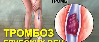
The formation of blood clots is a serious pathology that can threaten the patient’s life. A thrombus is formed from blood cells and is a dense clot. Blood clots can form in veins, arteries and the heart. The reason for this is various disorders of the cardiovascular system, bad habits, congenital pathologies of the body, etc. There is a high probability that the blood clot will break away from its permanent location and begin to travel through the blood vessels. If the clot enters the heart area, it can block the pulmonary artery, causing death. Many cases of sudden death are associated with undiagnosed thrombosis.
Most often, blood clots form in the lower extremities due to pathology of blood vessels or blood clotting disorders. Breaking off from the surface of the peripheral vein, the thrombus begins to move along the inferior vena cava and enters the right ventricle of the heart, and through it into the pulmonary artery. Thus, blockage of the branches of the pulmonary artery occurs.
A blood clot trap (a filter to catch blood clots) may be used to prevent this condition. A representative of such a trap is a vena cava filter. This is a special medical device that is implanted into the inferior vena cava. A clot trap creates an obstruction in the vein that stops large blood clots but does not interfere with normal blood flow.
A vena cava filter is a trap for blood clots, nothing more. It is not able to eliminate the very cause of blood clot formation. There are many medications available to treat thrombosis, but sometimes they are ineffective. The device is used in cases where conservative treatment does not bring the required result and measures must be taken to prevent pulmonary thromboembolism.
What vena cava filters are installed?
We use the most modern filter models, such as:
- permanent filter TrapEase (Cordis, USA),
- removable filters OptEase (Cordis, USA),
- Guenther Tulip (Cook, USA).
One of the latest innovations in the field of PE prevention is the ALN vena cava filter (ALN, France). This is the world's first vena cava filter that can be left in the vein for life or removed within up to 500 days. This is the longest possible removal period for removable vena cava filters.
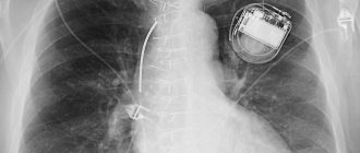
The vena cava filter is securely fixed in the vessel wall under the influence of body temperature. The filter has radiopaque markers made of gold rings built into the catheter introducer, which allow you to easily track the movement of the vena cava filter through tortuous vessels. The two-level structure of the filter allows you to effectively capture emboli of various sizes without disturbing the blood flow.
The presence of a vena cava filter in the vena cava does not affect the operation of the magnetic resonance imaging scanner, so a patient with a filter can undergo an MRI examination if necessary.
Blood to protect the body
Our organs and tissues are enriched with nutrients, remove waste, receive signals and transmit commands to control vital functions through the blood. Blood provides immunity and protection of the body not only from infections, but also from the occurrence of tumors and foreign tissues. This is not a complete list of the functions that blood performs.
From a chemical point of view, blood is a solution and suspension consisting of water and many water-soluble and insoluble substances and cells. But blood is also a living tissue, and it has unique properties. It can change viscosity, fluidity, gas and cellular composition depending on the situation.
One of the most important functions of blood is its ability to clot, the ability to quickly form clots called thrombi when necessary. On the other hand, after a certain time, blood enzymes can dissolve clots.
The main function of the blood coagulation system is protective. It allows you not to lose a significant amount of this unique fluid during injuries and wounds. A thrombotic “plug” helps prevent serious complications that arise from bleeding. But, unfortunately, with some congenital or, more often, acquired pathologies, thrombus formation in the body becomes excessive, and, as a rule, in the wrong place and at the wrong time.
How is a vena cava filter installed?
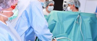
The procedure of implantation of a vena cava filter is currently a common endovascular intervention. The conductor with which the vena cava filter is implanted is inserted through the femoral access and the great saphenous vein, or into the jugular and subclavian veins under X-ray television control. The choice of access is determined by the intended location of the thrombus, since inserting a catheter through thrombosed veins is fraught with fragmentation of the thrombus with the development of pulmonary embolism. As a rule, the vena cava filter is installed at a level below the orifices of the renal veins.
Typically, the operation is performed in an X-ray operating room under short-term anesthesia (superficial anesthesia with propofol). During the implantation procedure, the patient does not feel any pain or discomfort.
Once the catheter reaches the desired level, the vena cava filter opens and the guidewire is removed. No stitches are placed on the skin. The duration of the placement procedure is usually about 1 hour. After implantation of the vena cava filter, a control x-ray is performed to confirm the correct installation and control its location.
We recommend limiting physical activity, preferably bed rest for 1–2 days after vena cava filter implantation. The patient is prescribed antibiotics and anticoagulants for 5–6 days.
How to install
Either the attending physician or the surgeon should tell the patient how to place blood clot traps. The patient must be fully aware of how the procedure will take place and how it will help their illness. At the Yusupov Hospital, specialists inform the patient in advance about all upcoming procedures. The patient receives complete information about the state of his health and can clarify any questions.
The installation of the vena cava filter is performed by an endovascular surgeon. This is a high-tech, minimally invasive operation that requires experience in vascular surgery. The operation is performed using local anesthesia, but the presence of an anesthesiologist is required.
At the beginning of the operation, catheterization of the vein is performed. Next, a vena cava filter is placed in the catheter and moved to a given point. The vena cava filter is in a special capsule at this time. When the clot trap reaches the required position, the capsule is removed. The vena cava filter straightens and grows into the walls of the vein, taking the correct position. The entire procedure is performed under the control of an ultrasound or X-ray machine.
It is very important to choose the correct size of the vena cava filter; it must match the diameter of the selected vein. A vena cava filter that is too small will not be able to anchor itself in the vein and may become dislodged. If the vena cava filter is large, it can damage the vessel walls.
The duration of the entire surgical intervention is on average half an hour. During the procedure, the patient does not experience pain. After the operation, the patient needs rest; bed rest must be observed. He is prescribed a course of antibiotics and medications with heparin.
What are the indications for installing vena cava filters?
The generally accepted indications for the installation of vena cava filters are:
- Deep vein thrombosis of the lower extremities with the presence of non-occlusive (floating) thrombi - a permanent vena cava filter is installed.
- Deep vein thrombosis of the legs with concomitant atrial fibrillation, coronary heart disease, heart failure, as well as in patients with cancer, especially in the later stages.
- Episodes of pulmonary embolism (PE) in the past in the presence of venous thrombosis of the lower extremities at the present time - a permanent vena cava filter is installed.
- Carrying out operations on the veins, laparoscopic interventions for thrombosis of the pelvic veins. At the time of surgery and the immediate postoperative period, a temporary vena cava filter is installed.
The vena cava filter itself does not affect the risk of blood clots, since it does not eliminate the cause of the blood clots. However, it prevents pulmonary embolism from a detached thrombus and reduces the risk of sudden death.
We install more than 20 different types of vena cava filters for patients, both temporary and permanent, from the world's leading manufacturers.
Preparing for surgery
- The doctor will give detailed instructions on how to prepare for the procedure.
- Do not eat or drink anything after midnight before your procedure.
- If you regularly take any important medications, you can take them with a sip of water.
- Do not smoke before and after the procedure. Smokers have a slower recovery after the procedure and are also more likely to have breathing problems during surgery. For this reason, if you are a smoker, you should quit smoking at least 2 weeks (preferably 6-8) before the procedure.
- Tell your doctor if you have any kidney problems or reactions to foods that contain iodine.
What restrictions do patients face after installing a vena cava filter?
When installing universal removable vena cava filters, the patient is under the dynamic supervision of an angiosurgeon or phlebologist for 6 months. If a vena cava filter is installed, taking warfarin is mandatory.
Once the risk of thromboembolism has been reduced, the temporary vena cava filter is removed using an intervention similar to inserting a filter. This operation is also performed in an X-ray operating room by an angiosurgeon under X-ray control.
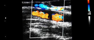
The fundamental decision to remove the vena cava filter is made after a repeat study of hemodynamics in the inferior vena cava system (including both limbs), when the dynamics of thrombosis resolution is assessed.
The installation of non-removable permanent vena cava filters, even in patients with advanced stages of cancer, is currently practiced relatively little, since a permanent vena cava filter, along with its positive role - preventing thromboembolism - causes obstruction of blood flow in the inferior vena cava basin and the formation of secondary venous insufficiency.
After installing a vena cava filter, the patient lives a normal life. We recommend taking indirect anticoagulants in maintenance dosages. In the presence of concomitant chronic venous insufficiency, it is advisable to undergo clinical observation with a visit to the doctor at least once a month, use phlebotonics and wear compression stockings. In addition, it is advisable to undergo a control duplex scan of the veins of the legs and the inferior vena cava.
A blood clot lurking against the wall
Under normal conditions, blood clots do not form inside blood vessels. However, if the vascular wall is injured or inflamed, the ability to clot blood increases, or blood flow through the vessels slows down, blood clots can form near the vessel wall (mural thrombus) or even completely block the vessel. A “fresh” thrombus slows down the blood flow even more, promoting the growth of the clot, which quickly increases, acquiring a loose and mobile “tail”. If part of the blood clot or all of it breaks off, then it becomes a dangerous “projectile” carried by the bloodstream throughout the human body.
What is the cost of installing a vena cava filter in Moscow?
We bring to your attention a price list from which you can find out the cost of installing vena cava filters at Euroonko:
- Implantation of a permanent vena cava filter made in the USA - 137,500 rubles.
- Implantation of a vena cava filter (made in the USA) with retrograde iliocavagraphy – RUB 179,300.
- Removal of a removable vena cava filter - 117,600 rub.
- Implantation of a removable vena cava filter made in the USA or France - 176,000 rubles.
Book a consultation 24 hours a day
+7+7+78
Our doctors
Drozdov Sergey Alexandrovich
Cardiovascular surgeon, phlebologist, Doctor of Medical Sciences
47 years of experience
Make an appointment
Malakhov Yuri Stanislavovich
Doctor - cardiovascular surgeon, phlebologist, Honored Doctor of the Russian Federation, Doctor of Medical Sciences, doctor of the highest category
Experience 36 years
Make an appointment
Removing a fixture
If the vena cava filter was initially installed as a temporary filter, then its removal is carried out by pulling on the conductor fixed under the skin.
If it is necessary to remove a permanent device, a catheter with a hook at the end is passed through the femoral vein. They grip the filter and then slide a cannula over it to cover its parts. After this, folded, it is removed from the vein. All these actions are carried out under intravenous anesthesia and visual control using ultrasound or radiography.
Device for removing the vena cava filter
When to choose temporary and when to choose permanent
Initially, it was assumed that the installed filters would be permanently in the vascular bed, and their removal was carried out only if serious complications developed. Since removable models have been developed, surgeons increasingly prefer their use.
Model of temporary vena cava filter 2014
Such a device can be installed for 2 - 3 months, that is, for as long as there is an increased risk of blood clots (for example, major surgery, trauma, heart valve replacement). After blood thinning has been achieved, or concomitant diseases have appeared that require removal of the filter, surgical removal of it from the vein can be performed.
Modern vena cava filters
If the period after implantation passes without complications, and the increased risk of blood clots in the deep venous network of the legs does not disappear, then the filter can be left.
Material and methods
A search was conducted for possible criteria for embolism in the CF using the Delphi method.
Moderators. The group of moderators consisted of surgeons from a clinic specializing in providing care to patients with venous thromboembolic complications.
Experts. 12 specialists were invited as experts who regularly treat patients with DVT and determine management tactics in cases where there are indications for CF implantation, as well as in the development of their occlusion.
Consensus. Achieved consensus on a particular criterion was considered to be the agreement of at least 75% of experts.
Rounds. The Delphi survey consisted of 3 rounds. In the first round, participants were encouraged to self-suggest criteria that they believed were indicative of embolism in the CF. In the second round, experts were asked to evaluate the results of the first round to express agreement or disagreement with them in order to highlight the most important criteria and exclude insignificant ones. At the third stage of the Delphi consensus, participants had to determine how many criteria, selected based on the results of voting in the second round, were necessary simultaneously to verify embolism in the CF.
Contraindications for thrombosis
As endovascular treatment technologies have advanced, insertion of a vena cava filter has become accepted as a routine method of preventing pulmonary embolism.
Despite this, anticoagulant therapy remains the main therapeutic factor for thrombosis, since it affects the cause of the disease, and surgery affects its consequences and complications.
In this case, contraindications to implantation may be identified:
- pathology of the inferior vena cava - excessive narrowing, inability to pass the catheter through nearby tissues and vessels, blockage above the renal segment;
- sepsis;
- malignant tumors;
- blood diseases.
In this regard, before referring the patient for the insertion of a vena cava filter, a complete angiographic examination is carried out to determine the patency of the vessels and select the required diameter of the device, and general clinical tests, ultrasound, and ECG are also prescribed.
Discussion
In order to develop criteria for the diagnosis of embolism in the CF, a modified Delphi method was used. Experts initially proposed 14 possible criteria for the differential diagnosis of embolism in the CF. It was possible to achieve consensus with a level of agreement of 75% or more on 4 criteria. The presence of any 2 of these criteria in one patient, according to the results of the third round, is confirmation of embolism in the CF. The remaining 10 criteria had a percentage level of agreement from 33.33 to 58.0%, which does not allow us to consider them as signs confirming the presence of CF occlusion of an embolic rather than thrombotic nature.
CF occlusion is currently considered one of the complications of the post-implantation CF period [8]. Typically, when analyzing the postimplantation period, the number of patients who developed IVC syndrome after CF implantation is reported. In this case, there is no distinction between filter occlusion of embolic origin and thrombosis in situ. Nevertheless, occlusion of embolic origin is fundamentally different from CF thrombosis in situ in that in the first case the filter fulfilled its task and prevented embolism in the pulmonary arteries, while in the second case, of course, we are talking about a complication of the procedure. However, the presence of differential criteria would be useful in assessing the effectiveness and safety of this common measure of surgical prevention of pulmonary embolism.
Due to the fact that such criteria are absent, it seems logical to develop them with the help of the expert opinion of specialists who regularly encounter such situations in clinical practice. The data obtained as a result of the Delphi consensus can become a starting point for further studies to analyze the use of CFs in terms of their effectiveness and safety, for a possible revision of the indications for CF implantation, as well as for the development of new recommendations for the treatment of venous thromboembolic complications.
Restrictions. Despite the high level of expert agreement regarding the developed criteria, the results obtained are based only on expert opinion, which may be erroneous or not correlate with clinical and instrumental data.
What is a device
Structurally, the filter is represented by a curved metal wire and resembles a bird's nest, umbrella spokes, and flower petals. The main characteristics of high-quality implants:
- resistance to destruction,
- tight fixation,
- should not interfere with blood flow,
- delay of large blood clots,
- easy installation and removal if necessary.
To do this, use steel, titanium or an alloy with nickel, and cover parts of the vena cava filters with a membrane that is saturated with Heparin.
The size is selected depending on the individual diameter of the vein chosen for installation. A catheter is used for delivery to the implantation area.
After it has reached the desired location, the structure opens and fixes itself due to elasticity or “tendrils.” Such filters are firmly connected to the venous wall and are installed on a permanent basis. Removal will require complex surgery. If a temporary option is provided, the vena cava filter is equipped with a conductor attached under the patient’s skin. This thread is used to remove the device after use.
We recommend reading the article about blockage of blood vessels in the legs. From it you will learn about the causes of the pathology and its symptoms, the methods of diagnosis and treatment used.
And here is more information about the prevention of thrombophlebitis.
How is the implantation procedure performed?
The procedure is performed under local anesthesia. Catheterization of the femoral artery or subclavian, jugular vein is carried out. Through the catheter, the guide delivers the filter to its place in a folded state. Under the control of an X-ray or ultrasound machine, the conductor is smoothly moved to the place where the filter is placed, freed from the media, straightening it and installing it at the desired point. It is important to assess the correspondence between the diameter of the device and the vein to avoid displacement or damage to the vessel walls. The intervention is painless and lasts up to 30 minutes.