There is very little knowledge about the capabilities of the human brain. Only its structure, ability to coordinate the work of the whole organism and its effect on overall well-being are known. For example, as a result of a disorder of blood flow in the cerebral artery, speech, coordination of movements, and thinking are disrupted, and paralysis occurs. All these are symptoms of a stroke. Brain disorders, in particular brain tumors, epilepsy, and Alzheimer's disease, have a worse impact on the duration and quality of life.
Timely and innovative diagnosis makes it possible to effectively treat diseases of any part of the brain.
Specialized methods
The following examination methods exist:
- Doppler ultrasound provides information about blood circulation in important vessels of the neck and brain. In this way, abnormalities of the vascular system are detected in the early stages. The effectiveness of the treatment is analyzed. Meanwhile, the day before you need to stop smoking and drinking caffeine. The above can affect vascular tone.
- Electroencephalography allows you to analyze the functional state of the brain and its irritability. In this case, even minor fluctuations are recorded. The information obtained is transferred to a special paper tape or converted into an image on a computer screen. This method makes it possible to diagnose and treat epilepsy, delayed mental and speech development, and detect the consequences of traumatic brain injuries.
- Echoencephalography diagnoses tumors and disorders of the brain structure, including after injuries. The device works by capturing a kind of echo, which is returned when ultrasonic waves are sent to the brain. The image is displayed on the screen.
- Rheoencephalography uses a weak high-frequency electric current to record fluctuations in the electrical resistance of tissues. This determines the condition of the vessels, their elasticity, blood filling and tone. The functioning of the arterial and venous systems of the brain is also established. Atherosclerosis, intracranial hypertension, subdural hematomas, and vascular dystonia are diagnosed. The effect of therapy for the listed diseases is assessed. The study is carried out using a rheograph apparatus with electrodes connected to it.
- Electroneuromyography. Using this method, brain biocurrents are recorded. The data obtained make it possible to diagnose dysfunctions of the peripheral nervous system and neuromuscular diseases. The procedure does not require lengthy and extensive preparation, and does not take much time, which makes it convenient and comfortable for those being examined.
- Neurosonography allows you to study the condition of babies from birth to 12 months. Ultrasound is used, so the procedure is safe. The equipment is highly accurate, as a result of which diseases are detected at the earliest stages, right up to the overgrowth of a large fontanel in the skull.
- Craniography. The examination is carried out using x-rays. Projections of the skull are made in profile and full face. This is how congenital or acquired bone abnormalities are detected. The value of craniography lies in the ability to quickly assess the presence of large fractures of the bones of the brain and facial skull. Craniography can be performed if there is a suspicion of a tumor of bone structures, brain structures, neuritis of the facial nerve, or if osteomyelitis is suspected.
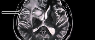
CT scan
Diagnosis is carried out by calculating the intensity of penetration of X-rays through brain tissue. A detailed image is displayed in cross section. The accuracy of the result is guaranteed even at low levels of radiation.
The examination is used if the patient suffers:
- pain in the head and neck area;
- fainting;
- dizziness;
- convulsions;
- speech and memory disorders;
- suffered a stroke;
- visual and auditory impairments.
The examination method under consideration is not applicable to pregnant women and children. If it is necessary to administer intravenously a contrast agent, the following contraindications are added:
- liver and kidney failure;
- heart defects;
- asthma;
- thyroid diseases;
- allergy to iodine;
- diabetes.
Before an examination using a contrast procedure, it is forbidden to consume food or liquid for 4 hours. Other cases do not require special preparation of patients. During the procedure, a person is moved on a moving table into a tomograph, where it is forbidden to move. At certain moments you will need to hold your breath.
In the absence of contraindications, the examination can be carried out as long as necessary to accurately establish the pathology.
Magnetic resonance imaging
MRI is very popular today. Thanks to the action of a magnetic field constantly maintained in the device, the condition of the skull is visualized. Hydrogen atoms present in the cells of the human body repulse the effects of electromagnetic waves. The data obtained is converted into images of brain tissue.
Diagnostics is effective for a wide range of pathologies: from diseases of the vascular system to tumors.
Contraindications to the examination include:
- mental disorders of the patient;
- acute pain syndrome or coma;
- metal and ferromagnetic pins, clips on blood vessels, implants in the patient’s body, permanent crowns on teeth;
- tattoos made with paint containing metallic particles.
The principle of operation is the same as that of computed tomography. The patient lies down on a moving table, the body is secured with straps, and sensory sensors are attached to the head. This is how the signal is sent and read. The table is sent to the tomograph. Duration - up to 40 minutes. The duration depends on the number of programs involved. The patient is required to lie still. The procedure is safe for children and adults.
Magnetic resonance angiography
The examinations are carried out according to the same rules as MRI. This is how pathologies of the vascular system are identified. The data is converted into a three-dimensional image of all brain vessels. The examination also allows the projection of thin sections of individual vessels and nerve trunks.
Positron emission tomography
This method examines the brain to record all ongoing functional processes. With its help, it is possible to distinguish a benign neoplasm from a malignant one in the early stages. The examination allows you to obtain information about abnormalities in the functioning of the brain, the consequences of injuries and bruises, and determine the condition of the organ after a stroke.
Patients are prohibited from eating 4-6 hours before. It is recommended to exclude foods containing protein the day before. The procedure involves intravenous administration of a radiopharmaceutical. The scan lasts 30-75 minutes.
Which study to choose if you have a headache?
Medical centers offer a wide range of head studies - ultrasound, EEG, REG, skull radiography, MRI, CT. “Which research method should I choose first?” - we asked this question to radiologist, candidate of medical sciences, Lidia Yuryevna Filippova. “First of all, with any type of headache, you first need to consult a neurologist. Headache can be a symptom of various diseases and conditions (hypertension, a consequence of injury, infections, pathology of vascular development, malignant and benign brain tumors, pathological changes in the cranial nerves, paranasal sinuses, etc.) At the appointment, a neurologist will conduct a detailed neurological examination and will prescribe the types of examinations he needs. If you don’t want to waste time and come to see a doctor with the results of examinations, it is optimal to choose MRI - it is a modern, safe (no ionizing radiation) and very informative examination method. Although it also has its contraindications (check when making an appointment). It is necessary to start with an MRI of the brain and cervical spine , because... In the vast majority of cases, headaches and dizziness are caused by pathology of the cervical spine (violation of the spinal axis, the presence of herniated intervertebral discs and marginal growths - osteophytes), which in turn affects the vessels of the neck (extravasal effects) and disrupts cerebral circulation. MRI of the brain, first of all, allows you to immediately exclude tumors, developmental anomalies, hemorrhages and cerebrovascular accidents, and with modern high-field tomographs with diffusion-weighted images, you can confidently exclude acute cerebrovascular accidents, even small ischemic foci. If you complain of pain in the neck, arm, numbness of the fingers, an MRI of the cervical spine is recommended to identify hernias and protrusions of intervertebral discs, their relationship with the nerve roots, assess the degree of narrowing of the spinal canal and the condition of the spinal cord. MRI in Domodedovo on Tekstilshchikov Building 2 performs MRI of the brain and cervical spine in a complex on a modern high-field MRI scanner. Based on the MRI results, the neurologist will receive a detailed description and conclusion from our specialist, which will allow him to quickly and accurately make a diagnosis and prescribe the correct comprehensive treatment. You will not only save money, but also start treatment on time, and most importantly, protect yourself from an incorrect diagnosis and, as a result, incorrect, and in some cases, life-threatening treatment.
How to choose an examination technique?
Ultrasound does not require special conditions for placement of equipment. This is the easiest way to diagnose. The purchase and installation of devices for CT, MRI or PET require considerable costs. In this regard, not all medical institutions can afford to carry out such procedures. For this reason, prices for these types of diagnostics will be high.
However, the popularity of the equipment and the price of diagnostics should not be the determining factors. First of all, you need to follow the recommendations of your doctor. The scope of application of the examination method should also be taken into account:
- PET detects a tumor, including a malignant one, with absolute accuracy, long before its manifestation.
- MRI is most effective in neurosurgery and neurology.
- CT is useful in detecting vascular damage and head injuries.
- From the point of view of the absence of ionization and X-ray radiation, MRI is the safest procedure. However, modern equipment for radiography and ultrasound significantly reduces the risk of gene mutations.
It is important not to forget about contraindications. Thus, PET and CT scans are strictly prohibited for pregnant women. MRI is used for expectant mothers if the potential benefit to the woman is higher than the possible risk to the baby.
Children require special preparation for the procedure. Parents should allegorically explain to their children the need to lie still. The youngest require anesthesia.
Only the attending physician can determine the need for a particular diagnosis. In some cases, different types of scanning may be required at the same time.