Causes of ventricular extrasystole
Sudden contraction of the ventricles is associated with the appearance of a focus of excitation in the Purkinje fibers or in the distal areas after the branching of the Githes bundle branches. The reasons causing the development of the disease can be divided into two blocks:
- Cardiac – causes associated with heart disease:
- ischemia;
myocardial infarction;
- cardiomyopathy;
- cardiosclerosis;
- heart defects.
- Extracardiac or extracardiac – functional factors:
- drug overdose;
infectious diseases;
- consumption of tonic and energy drinks;
- physical exercise;
- stress, increased emotional tension;
- abuse of bad habits.
General information
The factors that caused the development of the disease can be of physiological and pathological origin. An increase in the tone of the sympathetic-adrenal system leads to an increase in the occurrence of extrasystoles. Physiological factors influencing this tone include the consumption of coffee, tea, alcohol, stress and nicotine addiction. There are a number of diseases that lead to the formation of extrasystole:
- cardiac ischemia;
- myocarditis;
- cardiomyopathy;
- heart failure;
- pericarditis;
- hypertonic disease;
- osteochondrosis of the cervical spine;
- prolapse of the mitral valve leaflets;
- cardiopsychoneurosis.
There is a certain connection between the patient’s age, time of day and the frequency of extrasystoles. Thus, more often the ventricular type is present in people over 45 years of age. Dependence on circadian biorhythms is manifested in the registration of extraordinary heart contractions, more in the morning hours.
Ventricular extrasystole threatens the patient’s life. Its formation increases the risk of sudden cardiac arrest or ventricular fibrillation.
Symptoms
Ventricular extrasystole can be asymptomatic or have a pronounced clinical picture:
- interruptions in the functioning of the heart - first rapid heartbeat, then freezing;
- dizziness;
- weakness;
- unpleasant, painful sensations in the heart;
- pulsation of the neck veins;
- fast fatiguability;
- reduced performance;
- lack of air;
- shortness of breath.
If you notice similar symptoms or feel unwell, contact a specialist immediately.
Modern approach to the diagnosis and treatment of ventricular extrasystole
The etiology of PVCs is multifactorial, idiopathic (not associated with any organic pathology), PVCs against the background of organic, dysplastic, sclerotic, scar changes, dilatation, hypertrophy, ventricular myocardium. Patients with PVCs complain of interruptions in the functioning of the heart, sometimes associated general weakness, dizziness and even fainting (syncope). In combination with reduced cardiac ejection fraction and previous myocardial infarction, PVC is a predictor of sudden death in patients.
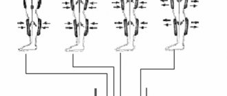
The main method for diagnosing PVCs is electrocardiography (ECG), which allows not only to establish a diagnosis, but also, with a fairly high degree of probability, to determine the localization of ectopia, and when recording it daily, the absolute number of extrasystoles. There are a number of classifications of PVCs, the most used of them is the classification of B. Lown and M. Wolf (1971), according to which ventricular extrasystole is divided into five gradations: 0 – absence of PVCs, 1 – rare monotopic up to 30 per hour, 2 – frequent monotopic more 30 per hour, 3 – polymorphic PVCs, 4 – A paired PVCs, B – volley PVCs, runs of ventricular tachycardia (3 or more complexes), 5 – early PVCs R on T. However, later, according to some studies, it was found that early extrasystoles do not carry such a large prognostic load, and a revised classification was adopted (B.Lown and M.Wolf (1971), as modified by M.Ryan et al. (1975)): absence of PVCs during 24 hours of monitor observation - 0; no more than 30 PVCs in any hour of monitoring - I; more than 30 ectopic ventricular complexes during any hour of monitoring - II; polymorphic PVCs - III; monomorphic paired PVCs - IV-A; polymorphic paired PVCs - IV-B; ventricular tachycardia (VT) - three or more consecutive VVCs with a frequency of more than 100 per minute) - V. Functional classes 3-5 are classified by the authors as extrasystole of high gradations. Patients with rare asymptomatic PVCs do not require antiarrhythmic therapy (AAT). Frequent monotopic PVCs (4, 5 grades) without an organic nature (idiopathic) are subject to AAT, while drugs of class I according to the Vaughan Williams EM 1984 classification (Etatsizin, Propafenone) are most often used, with class III ineffective (d-Sotalol, Amiodarone). In the absence of an effect from adequate drug therapy and the number of extrasystoles is 10 thousand per day or more, or when PVCs return after discontinuation of antiarrhythmics, surgical treatment is indicated, catheter radiofrequency destruction of the extrasystolic focus (Fig. 1 - 3), the effectiveness of which is more than 90%. PVCs associated with electrolyte disturbances (hypokalemia) require correction of these disturbances.
PVCs, most often polytopic, against the background of organic changes in the ventricular myocardium, stenosing atherosclerosis of the coronary arteries, in the presence of post-infarction scars, chronic cardiac aneurysm, severe hypertrophy or dilatation of the myocardium, require, in addition to routine echocardiography, a study of the degree and nature of coronary lesions (coronary angiography) , in order to determine indications for myocardial revascularization (angioplasty and stenting, coronary artery bypass grafting (CABG)), for plastic surgery (aneurymectomy). These patients are contraindicated in the use of class I antiarrhythmic drugs, since according to multicenter studies (CAST-I and CAST-II), they significantly increase the risk of sudden death due to the occurrence of life-threatening cardiac arrhythmias. Such patients are prescribed AAT using β-blockers (mortality reduction by 35%, Cannon DS, Prystowsky EN, 1999) and class III drugs (Amiodarone, mortality reduction by more than 30%, CAMIAT, EMIAT studies; or d-Sotalol) with ECG monitoring of the duration of the QT interval, prolongation of which by more than 500 ms can lead to the development of fusiform ventricular tachycardia (VT) “torsades de pointes”, which can transform into ventricular fibrillation (VF). In this case, the proper QT interval is determined by the Bazet formula: QTd = k x √RR, where the “k” index for women is 0.40, and for men 0.37. In addition, patients who have had a myocardial infarction, 4 weeks or 3 months after CABG, with a low (less than 35%) left ventricular ejection fraction are advised to undergo implantation of a cardioverter-defibrillator to prevent fatal arrhythmias (VT, VF).
Thus, ventricular extrasystole is a very serious problem; patients suffering from it require careful examination, constant monitoring, adequate antiarrhythmic therapy and correction of the pathology underlying the occurrence of ectopia. All these patients should be consulted by an arrhythmologist.
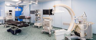
Currently, in the Clinical Hospital No. 1 of the Administration of the President of the Russian Federation, Moscow, on the basis of the department of x-ray methods of diagnosis and treatment, an arrhythmology service is successfully operating, which has the most modern equipment and consumables from the world's best manufacturing companies. The department performs the full range of high-tech studies and operations for any rhythm disturbances.
Mezentsev P.V., Ph.D. Zakaryan N.V.
Diagnosis of ventricular extrasystole
Diagnosis of VES includes laboratory and instrumental studies. First, the doctor interviews the patient, conducts an examination, measures blood pressure and, based on the results, gives a referral for an ECG and Holter monitoring. Also used when diagnosing the disease:
- ECHO;
- MRI;
- EFI;
- angiography;
- load tests.
Timely diagnosis of VES will allow you to detect the disease in time, stop it and avoid complications.
Prevention
Detection of ventricular extrasystole on an ECG does not always mean serious problems; in a mild form, the disease does not require special treatment. It is enough to follow the doctor’s recommendations, undergo an examination by a cardiologist, avoid alcohol, energy or tonic drinks, and stop smoking. Regulate your daily routine, get enough sleep, walk in the fresh air, do therapeutic exercises, avoid excessive physical activity and stressful situations.
In cases where PVC is accompanied by heart disease, it is necessary to undergo examination by a cardiologist. The cardiology center of the Federal Scientific and Clinical Center of the Federal Medical and Biological Agency has developed several heart research programs. You can get acquainted with them by following the link. Do not under any circumstances stop prescribed medications on your own, do not disrupt your medication regimen, and do not treat yourself with traditional medicine.
Reasons for development
Irregularities and heart diseases are the main reasons why PVCs develop. Also, ventricular arrhythmia can be triggered by heavy physical work, chronic stress and other negative effects on the body.
From cardiac pathologies:
| Heart failure | Negative changes in the muscle tissue of the heart muscle, leading to disruption of the inflow and outflow of blood. This is fraught with insufficient blood supply to organs and tissues, which subsequently causes oxygen starvation, acidosis and other metabolic changes. |
| Coronary heart disease (CHD) | This is damage to the heart muscle due to impaired coronary circulation. IHD can be acute (myocardial infarction) or chronic (with periodic attacks of angina). |
| Cardiomyopathy | Primary myocardial damage, leading to heart failure, atypical beats and heart enlargement. |
| Heart disease | Defect in the structure of the heart and/or major outflow vessels. Heart disease can be congenital or acquired. |
| Myocarditis | An inflammatory process in the heart muscle that disrupts impulse conduction, excitability and contractility of the myocardium. |
Taking certain medications (incorrect dosage, self-medication) can also affect the functioning of the heart:
- Ventricular extrasystole
| Diuretics | Medicines in this group increase the rate of urine production and excretion. This can provoke excessive excretion of the “heart” element - potassium, which is involved in the formation of the impulse. |
| Cardiac glycosides | The drugs are widely used in cardiology (they lead to a decrease in heart rate and an increase in the force of myocardial contraction), but in some cases they cause side effects in the form of arrhythmia, tachycardia, atrial fibrillation and ventricular fibrillation. |
| Drugs used for heart blocks (M-anticholinergics, sympathomimetics) | The side effects of the drugs manifest themselves in the form of stimulation of the central nervous system, increased blood pressure, which directly affects heart rhythm. |
Also, the development of PVCs can be affected by other pathologies not associated with disruption of the cardiovascular system:
- Diabetes mellitus type 2 . One of the serious complications of the disease associated with carbohydrate imbalance is diabetic autonomic neuropathy, which affects nerve fibers. In the future, this leads to a change in the functioning of the heart, which “automatically” causes arrhythmia.
- Hyperfunction of the thyroid gland (moderate and severe thyrotoxicosis). In medicine, there is such a concept as “thyrotoxic heart,” which is characterized as a complex of cardiac disorders - hyperfunction, cardiosclerosis, heart failure, extrasystole.
- In diseases of the adrenal glands, there is an increased production of aldosterone, which in turn leads to hypertension and metabolic disorders, which is interconnected with the work of the myocardium.
Ventricular extrasystole is not of an organic nature (when there are no concomitant cardiac diseases), caused by a provoking factor, often has a functional form. If you remove the negative aspect, then in many cases the rhythm returns to normal.
Functional factors of ventricular extrasystole:
- Electrolyte imbalance (reduction or excess of potassium, calcium and sodium in the blood). The main reasons for the development of the condition are changes in urination (rapid production or vice versa, urinary retention), malnutrition, post-traumatic and postoperative conditions, liver damage, and surgery on the small intestine.
- Substance abuse (smoking, alcohol and drug addiction). This leads to tachycardia, changes in material metabolism and disruption of myocardial nutrition.
- Disorders of the autonomic nervous system due to somatotrophic changes (neuroses, psychoses, panic attacks) and damage to subcortical structures (occurs with brain injuries and pathologies of the central nervous system). This directly affects the functioning of the heart and also provokes surges in blood pressure.
Ventricular extrasystoles disrupt the entire heart rhythm. Pathological impulses over time have a negative effect on the myocardium and the body as a whole.
How to treat ventricular extrasystole
When treating ventricular extrasystole, classical drug therapy or surgery is used. The treatment regimen is always determined by a cardiologist.
In our center of the Federal Scientific and Clinical Center of the Federal Medical and Biological Agency there is a therapeutic department in which the patient can undergo all the necessary heart examinations and tests under the supervision of specialists.
The surgical department employs doctors with many years of experience who perform heart surgeries of any complexity. In the case of ventricular extrasystole, radiofrequency catheter ablation may be proposed - a targeted effect of the electrode on areas with impaired conductivity.


![Rice. 1. ACCOMPLISH study: the effect of the combination of ACE inhibitor* amlodipine and ACE inhibitor hydrochlorothiazide on the risk of primary endpoint events (CVE) [14]](https://expert35.ru/wp-content/uploads/ris-1-issledovanie-accomplish-vliyanie-kombinacii-iapf-amlodipin-i-iapf-330x140.jpg)


