Magazine "Medical Council" No. 12
/2021
DOI:
10.21518/2079-701X-2021-12-353-359
A.G. Syrkasheva
, ORCID: 0000-0002-7150-2230
N.V.
Dolgushina , ORCID: 0000-0003-1116-138X
National Medical Research Center for Obstetrics, Gynecology and Perinatology named after Academician V.I. Kulakova; 117997, Russia, Moscow, st. Academician Oparina, 4
Introduction
. Infertility, i.e. failure to achieve clinical pregnancy within 12 months. regular sexual activity without contraception is a pressing medical problem and affects up to 15–25% of married couples in Western countries.
Target
. To evaluate the effectiveness of prophylactic administration of antioxidants in preparation for assisted reproductive technology (ART) cycles, depending on the level of anthropogenic chemicals in the patient’s body.
Materials and methods
. The randomized clinical trial included 144 patients with infertility who applied for ART. Before treatment with ART methods, all patients had the level of anthropogenic chemicals (ACS) in the blood determined by mass spectrometry. The concentration of the following substances was determined: mercury, cadmium, lead, bisphenol A. To assess the total level of chemical substances, a conventional scale, developed earlier, was developed. The patients were divided into groups depending on the level of AChB: group 1 consisted of 72 patients with a high level of AChB (5 points or more), group 2 consisted of 72 patients with a low level of AChB. Antioxidant therapy in the experimental group was carried out for 2 months. before ART. Coenzyme Q10 300 mg/day orally, eicosapentaenoic acid 300 mg/day orally, and docosahexaenoic acid 200 mg/day orally were used as antioxidant therapy. All patients in the control group did not take antioxidant drugs for at least 6 months. before entering the ART cycle.
results
. When assessing the clinical outcomes of ART cycles, a positive effect of antioxidant therapy was noted in both patients with high levels of AChB and patients with low levels of AChB; as a result, in the group of patients receiving antioxidant therapy, the chances of pregnancy were 2.3 times higher compared to control group. The number of patients treated was 5 both for the overall group of patients and for subgroups depending on the level of ACV.
Conclusion
. The results obtained allow us to recommend the use of antioxidant therapy to prepare patients for ART programs.
For quotation:
Syrkasheva A.G., Dolgushina N.V. The role of antioxidant therapy in increasing the effectiveness of assisted reproductive technology programs. Medical advice. 2021;(12):353-359. https://doi.org/10.21518/2079-701X-2021-12-353-359
Conflict of interest:
The authors declare no conflict of interest.
The role of antioxidant therapy in enhancing the effectiveness of assisted reproductive technology programs
Anastasia G. Syrkasheva
, ORCID: 0000-0002-7150-2230
Nataliya V. Dolgushina
, ORCID: 0000-0003-1116-138X
Kulakov National Medical Research Center for Obstetrics, Gynecology and Perinatology, 4, Academician Oparin St., Moscow, 117997, Russia
Introduction
. Infertility, ie the inability to achieve a clinical pregnancy within 12 months of a regular sexual life without contraception, is a current medical problem and affects up to 15-25% of married couples in Western countries.
Objective
. To evaluate the efficacy of prophylactic prescription of antioxidants in preparation for cycles of assisted reproductive technology (ART) depending on the level of anthropogenic chemicals in the patient's body.
Materials and methods
. A randomized clinical trial included 144 patients with infertility who applied for ART. Prior to ART treatment, all patients were determined the level of anthropogenic chemical substances (ACS) in the blood by mass spectrometry. The concentration of the following substances was determined: mercury, cadmium, lead, and bisphenol A. The patients were divided into groups depending on the level of ACS: group 1 consisted of 72 patients with high level of ACS (5 points or more), group 2 consisted of 72 patients with low level of ACS. Antioxidant therapy in the experimental group was performed for 2 months before ART. Coenzyme Q10 300 mg/day orally, eicosapentaenoic acid 300 mg/day orally, and docosahexaenoic acid 200 mg/day orally were used as antioxidant therapy. All patients in the control group did not take antioxidant medications for at least 6 months before entering the ART cycle.
Results
. When evaluating the clinical outcomes of ART cycles, a positive effect of antioxidant therapy was noted in both patients with high and low levels of ACS; as a result, the chances of pregnancy were 2.3 times higher in the group of patients who received antioxidant therapy compared to the control group. The number of patients treated was 5 for both the total patient group and the subgroups depending on the level of ACS.
Conclusion
. The results obtained allow us to recommend the prescription of antioxidant therapy to prepare patients for ART programs.
For citation:
Syrkasheva AG, Dolgushina NV The role of antioxidant therapy in enhancing the effectiveness of assisted reproductive technology programs. Meditsinskiy sovet = Medical Council. 2021;(12):353-359. (In Russ.) https://doi.org/10.21518/2079-701X-2021-12-353-359
Conflict of interest:
the authors declare no conflict of interest.
Introduction
Infertility, i.e. failure to achieve clinical pregnancy within 12 months. regular sexual activity without contraception is a pressing medical problem and affects up to 15–25% of married couples in Western countries [1]. According to some reports, the number of married couples with impaired fertility may be twice as high, given the difficulties in carrying a pregnancy to term [2].
Over the past decades, the need for the use of assisted reproduction methods has increased. For example, in the USA in 1995, 60 thousand cycles of assisted reproductive technologies (ART) were performed, and in 2015 – 209 thousand cycles. According to the Russian Association of Human Reproduction (RAHR), in 2000, 6 thousand ART cycles were performed in Russia, and in 2021 - almost 146 thousand cycles. However, despite the scientific achievements of recent decades, the effectiveness of ART programs does not exceed 30–35% and does not tend to increase.
The high prevalence of infertility and the growing need for ART methods motivate to identify modifiable factors affecting reproductive health. In recent years, much attention from the scientific community has been paid to lifestyle modification and limiting contact with harmful environmental factors as part of preconception preparation [3, 4].
One of the modern trends in reproductive medicine is the prescription of oral antioxidants (mainly in the form of dietary supplements) to patients planning independent pregnancy or ART cycles. However, there is insufficient data on the effectiveness of these drugs in the scientific literature. One potential mechanism of action of antioxidant drugs may be a reduction in oxidative stress. Oxidative stress is the main pathogenetic mechanism of the negative impact of harmful environmental factors on the human body [5]. Increased levels of anthropogenic chemicals are associated with fertility disorders, as the reproductive system is vulnerable to external factors [6].
Purpose of the study
– to evaluate the effectiveness of prophylactic administration of antioxidants to prepare for ART cycles depending on the level of anthropogenic chemicals in the patient’s body.
Antioxidants in the treatment of cerebrovascular diseases
Currently, cerebrovascular pathology ranks second among the main causes of mortality, second only to heart disease and already ahead of mortality from tumors of all locations. Cerebrovascular pathology is the leading cause of disability in the population and, therefore, represents one of the most important medical and social problems.
Today, about 9 million people worldwide suffer from cerebrovascular diseases. The leading role among these diseases is occupied by strokes, affecting from 5.6 to 6.6 million people annually and claiming 4.6 million lives. According to the World Health Organization, the incidence of stroke ranges from 1.5 to 7.4 per 1000 people. Thus, in the United States, a cerebral stroke occurs every 53 seconds.
In the Russian Federation and CIS countries, there is a progressive increase in the incidence of this pathology: approximately every 1.5 minutes, one of the Russians develops a stroke for the first time. The incidence of stroke in Russia is 450,000 cases per year: in Moscow alone, the number of acute strokes ranges from 100 to 120 cases per day. The overall mortality rate from stroke in 2001 was 1.28 per 1000 people (men - 1.15, women - 1.38). The mortality rate from stroke in our country is one of the highest in the world: in 2000, the standardized rate was 319.8 per 100,000 people. In terms of mortality rates, Russia ranks second, second only to Bulgaria. Mortality in the acute stage of all types of stroke is approximately 35%, increasing by another 12–15% by the end of the first year. Along with high mortality, the consequences of strokes are also socially significant - the development of disability with loss of ability to work. Disability after a stroke ranks first among all causes of primary disability, since less than 20% of survivors return to their previous social and work activities. In addition, enormous damage is caused to the economy, taking into account the costs of treatment, medical rehabilitation, and losses in production. In the USA, material costs for strokes range from 7.5 to 11.2 million dollars per year, costs per patient, taking into account the need for long-term treatment and social rehabilitation, range from 55 to 73 thousand dollars per year.
The ratio between ischemic and hemorrhagic stroke was previously 5:1. Registry data from 2001 showed that in Russia ischemic strokes amounted to 79.8%, intracerebral hemorrhages - 16.8%, subarachnoid hemorrhages - 3.4%.
In Russia, up to 100,000 new cases of cerebral hemorrhages are registered annually. The incidence of hemorrhagic stroke is higher in men, while the mortality rate is higher in women. According to a number of authors, mortality from cerebral hemorrhage varies from 38 to 93%, with 15–35% of patients dying within a month from the moment of illness, half of them die within the first three days. Only 10% of patients by the end of the first month and 20% after six months can care for themselves; 25–40% of patients have a moderate degree of disability, 35–55% have severe disability.
The epidemiological and demographic situation in the world regarding cerebrovascular pathology is currently characterized by the widespread prevalence of this type of pathology, the “aging” of the population and the increase in the frequency of progressive cerebrovascular diseases, the “rejuvenation” of strokes due to the increase in the number of extreme factors and impacts (A. A. Mikhailenko and co-authors, 1996; A. A. Skoromets, 1999). In a large number of people over the age of 50, the processes of so-called “normal aging” are quickly replaced by pathological changes associated primarily with insufficiency of cerebral blood flow due to atherosclerotic damage to the vessels supplying blood to the brain, with changes in the rheological properties of the blood, leading to dysregulation and decreased neurotransmitter activity. Clinically, these neurotransmitter and morphological dysregulations are manifested by severe symptom complexes of acute and/or chronic cerebral ischemia, requiring constant and effective correction.
The number of patients with symptoms of chronic cerebral ischemia in our country is growing as steadily as the number of patients with acute cerebrovascular accidents, amounting to at least 700 per 100,000 people. While our country currently has statistics on acute strokes, albeit not in full, there are no reliable statistics on the number of patients with chronic cerebral ischemia. These are mainly outpatient patients; visiting a clinic is often difficult for them; Often they are given complex diagnoses, while cerebrovascular pathology is not taken into account or is classified as a complication, which makes it difficult to obtain objective data. The shortage of qualified neurologists in outpatient clinics also often leads to incorrect interpretation of this diagnosis.
Pathomorphological disorders in patients with acute and chronic cerebral ischemia are based on a variety of pathogenetic factors, such as atherosclerosis, arterial hypertension, as well as their combinations, cardiac pathology, changes in the condition of the spine with compression of the vertebral arteries, hormonal disorders leading to changes in the coagulation system blood, other types of disorders of the hemostatic system and physicochemical properties of blood, leading to the formation of functional and morphological ischemic disorders.
The most common causes of the formation of clinical manifestations of cerebral ischemia are atherosclerotic stenotic and occlusive lesions of the main arteries of the head; heart diseases, which include primarily coronary heart disease with symptoms of atrial fibrillation and a high risk of microembolization into intracerebral vessels. Atherosclerosis is a systemic vascular disease that leads to infiltration of the intima of the arteries with cholesterol coming from the blood. In the development of atherosclerosis, hereditary predisposition and constitutional characteristics play a role. However, the main reason for the wide spread of atherosclerosis in recent years is the functional effects on higher nervous activity of a person, which can be classified as negative manifestations of urbanization in the conditions of scientific and technological progress. They lead to long-term and systematic neuropsychic stress. The development of atherosclerosis is promoted by physical inactivity and hypokinesia (work without physical exertion, limited walking, passive rest), hypoxia (urban air pollution), increased exposure to external electromagnetic potential, the negative impact of noise and the pace of city life, insufficient sleep and excess calorie content of food (taking into account hypokinesia). The widespread use of smoking in recent years as a factor contributing to the development of vasospasms in various vascular systems is also of known importance. In this regard, in recent years, about a contingent of patients with atherosclerosis and arterial hypertension, in particular, from 50 to 60% of cases of cerebral vascular diseases occur between the ages of 50 and 60 years. At the same time, cerebral atherosclerosis takes first place compared to arterial hypertension. Four of the factors noted above are of leading importance in the development of vascular cerebral pathology, in particular atherosclerosis: neuropsychic stress, hypokinesia, physical inactivity and excess calorie intake. As a result of their influence, overexcitation of the cerebral cortex and the hypothalamic-pituitary-adrenal system occurs, increased release of catecholamines, disruption of all types of metabolism, especially in the walls of blood vessels, and sometimes increased blood pressure.
The study of the causes of morbidity and mortality in vascular diseases of the nervous system has led to the establishment of risk factors that play a contributing role in the development of cerebral vascular accidents. These factors include: arterial hypertension, vascular hypotension, obesity (overweight), hypercholesterolemia (especially in young and middle-aged people), smoking, alcohol abuse, family history, coronary atherosclerosis, diabetes mellitus, endocrine pathology, mineral metabolism disorders (cervical osteochondrosis), living in areas with sharp fluctuations in meteorological factors, work with high intellectual stress.
Hemorrhagic stroke, also characterized by a severe secondary ischemic cascade, most often occurs as a complication of arterial hypertension (60% of cases). The development of degenerative changes (lipohyalinosis, fibrinoid necrosis) in small perforating arteries of the brain and the formation of microaneurysms against the background of arterial hypertension are the most important prerequisites for the occurrence of hypertensive intracerebral hemorrhage, and hemorrhage develops more often in patients with severe or moderate arterial hypertension than in patients with “mild” » arterial hypertension. Pathogenetically, intracerebral hemorrhages develop due to rupture of a vessel or through diapedesis. The next most common etiological factor for cerebral hemorrhage is rupture of an arteriovenous malformation, hemorrhage from ruptured aneurysms (10–12% of cases). Occurring more often in old age, cerebral amyloid angiopathy, which is formed due to the deposition of abnormal amyloid protein in the tunica media and adventitia of small cortical arteries and arterioles, contributes to the occurrence of miliary aneurysms and fibrinoid necrosis of the affected vessels, which can rupture when blood pressure rises, causing intracerebral hemorrhage in 10 % of cases. Such hematomas are often multiple. Long-term use of anticoagulants in 8–10% of cases leads to intracerebral hemorrhage, especially when hypocoagulation is achieved, i.e., a decrease in the prothrombin index to 40% or an increase in the international normalizing coefficient to 5. Brain tumors or brain metastases are complicated by hemorrhages in them in 6 -8% of cases. Up to 20% are other causes, such as hemophilia, thrombocytopenia, leukemia, hemorrhagic diathesis, arteritis, thrombosis of intracranial veins, alcohol and drug abuse, coagulopathy, vasculitis.
The mechanism of development of hypoxia, which is a discrepancy between tissue demand for oxygen and its delivery, is the same for any form of cerebrovascular pathology. It is associated primarily with impaired oxidation of substrates in body tissues as a result of difficulty or blockade of electron transport in the mitochondrial respiratory chain, which leads to damage to lysosome membranes with the release of utilitarian enzymes into the intercellular space.
Stress, or more precisely distress according to Selye’s theory, is a mechanism of nonspecific adaptation to the changing conditions of the organism’s environment.
At the initial stage of oxygen starvation in mitochondria, the rate of aerobic oxidation and oxidative phosphorylation decreases, which leads to a decrease in protein synthesis and gene expression, a decrease in the amount of adenosine triphosphate (ATP), an increase in adenosine diphosphate (ADP) and adenosine monophosphate (AMP); the ATP/ADP+AMP ratio decreases. With a further decrease in cerebral blood flow, the enzyme phosphofructokinase (PFK) is activated, anaerobic glycolysis is enhanced, and then a final transition to anaerobic respiration is noted, which adapts the cell to hypoxia, but glycogen reserves are depleted. This, in turn, entails the accumulation of under-oxidized lactate, a decrease in pyruvate with the development of lactic acidosis - up to the development of cerebral edema.
At the same time, the activity of lactate dehydrogenase increases and the activity of succinate dehydrogenase, which supplies electrons to the respiratory chain of mitochondria, decreases, which indicates a disruption in the processes of energy formation in the ischemic brain. Under such conditions, anaerobic glycolysis does not occur, which leads to severe energy deficiency. At the final level, destabilization of cell membranes occurs, disruption of the functioning of ion channels, damage to the potassium-sodium pump, potassium (an excitatory neurotransmitter) leaves the cell, which makes it less excitable, and sodium enters the cell in excess, followed by sodium along the osmotic gradient and enters the cell Excessive amounts of water leaving the interstitium accumulate, which leads to cell hyperhydration, cloudy swelling, and then balloon degeneration. The most important role in this process belongs to glutamate receptors.
Oxidative stress, closely associated with the ischemic cascade, occurs when glutamate receptors are excited and consists of excessive accumulation of free radicals, activation of lipid peroxidation and excessive intracellular accumulation of their products. The reactions of oxidative stress and the ischemic cascade interact and potentiate each other.
Free radicals (these are molecules with an unpaired electron) are highly reactive forms of oxygen, hydrogen peroxide, aldehydes formed under hypoxic conditions, with incomplete reduction of oxygen, changing the functional properties of a number of enzymes, carbohydrates, proteins, including deoxyribonucleic acid (DNA) and ribonucleic acid (RNA), as a result the cell loses its functions, abnormal proteins appear and, in addition to the direct damaging effect, secondary destructive processes are stimulated. Oxygen for any cell, especially for a neuron, is the main energy acceptor in the mitochondrial respiratory chain. By binding to the iron atom of cytochrome oxidase, the oxygen molecule undergoes four-electron reduction to form water. The main stable form of oxygen is “triplet” oxygen, in the molecule of which both unpaired electrons are parallel and their valences (spins) are directed in the same direction. Oxygen, in the molecule of which the valences are directed in different directions, is called singlet, it is unstable and toxic for biological substances. Free radicals are unstable and tend to transform into stable compounds by pairing a free radical, tearing off an atom, most often hydrogen, from another compound and attaching it to itself.
Along with the processes of free radical oxidation, stable antioxidant radicals are produced in biological objects, which are capable of abstracting hydrogen atoms only from special molecules that have weakly bound hydrogen atoms. This class of chemical compounds is called antioxidants, since their mechanism of action is based on inhibition of free radical processes in tissues, which inhibits the development of destructive changes and inactivates oxidative stress reactions. Changes in the structure and function of substrates under conditions of ischemia and stress depend on the ratio of the activity of free radicals and antioxidants.
It should be noted that the pathophysiological mechanisms of the emergence and progression of oxidative stress in patients with any form of cerebrovascular pathology are the same and are characteristic of both patients with ischemic and hemorrhagic stroke and patients with chronic forms of cerebrovascular insufficiency. Chronic cerebral ischemia is a disease that progresses stepwise against the background of repeated episodes of dyscirculation, leading to an increase in brain hypoxia.
Treatment of cerebral stroke consists of general and specific methods. The first include measures to ensure adequate oxygenation, correction of blood pressure, relief of complications, possible seizures, monitoring the condition of vital organs, patient care measures, as well as the use of specific therapy methods that stimulate the protective mechanisms of brain tissue in conditions of acute ischemia and hypoxia . The same applies to the processes of correction of chronic forms of cerebral circulatory disorders.
One of the most promising methods of nonspecific therapy for cerebral stroke and chronic forms of cerebral circulatory disorders is currently the use of antioxidants, which are specific correctors of brain energy metabolism, acting specifically under conditions of ischemia and hypoxia.
The body has a physiological antioxidant system that maintains oxidative-antioxidant balance both in liquid media (blood, lymph, intracellular and intercellular fluid) and in the structural elements of the cell (plasmic, endoplasmic, mitochondrial, cell membranes). Enzymatic antioxidants include: superoxide dismutase, which inactivates the superoxide radical inside the cell; catalase, which decomposes intracellular hydrogen peroxide; glutathione dehydroascorbate reductase, some other peroxidases.
Non-enzymatic antioxidants include vitamins C, E, K, glucose, ubiquinones, phenylalanine, transferrin, haptoglobin, tryptophan, ceruloplasmin, carotenoids. Biological and chemically synthesized antioxidants are divided into fat-soluble and water-soluble. The former are localized where the target substrates for attack by free radicals and peroxides are located, the most vulnerable biological structures to peroxidation processes, which include primarily biological membranes, blood lipoproteins, and the main targets in them are unsaturated fatty acids. The most significant fat-soluble antioxidant is α-tocopherol; it interacts with the hydroxyl radical OH and has an inhibitory effect on singlet oxygen, preserving the activity of membrane-bound enzymes. α-tocopherol is not synthesized in the body; it belongs to the group of vitamins (vitamin E), is a universal fat-soluble antioxidant and a natural immunomodulator, normalizing the indicators of cellular and humoral immunity. Among the water-soluble antioxidants, the most important are glutathione, which plays a key role in protecting cells from toxic oxygen intermediates, and the ascorbic acid system, which is especially important for the antioxidant protection of the brain. It should be noted that antioxidants supplied in food also take part in the fight against oxidative stress: minerals (selenium, magnesium, copper compounds), some amino acids, flavonoids (plant polyphenols). However, their role is reduced to a minimum if we take into account that the diet of a modern person is dominated by refined and processed foods that lack natural qualities (even if plant products predominate in the diet), which is the cause of chronic deficiency of antioxidants in the human body.
The most adequate synergist and almost ubiquitous companion of ascorbic acid is the system of phenolic compounds. It is found in all plant living organisms, making up 1–2% of biomass or more, and performs various biological functions.
The antioxidant properties of phenols are associated with the presence in their structure of weak phenolic hydroxyl groups, which easily give up their hydrogen atom when interacting with free radicals and act as free radical traps, turning into low-active phenoxyl radicals. The greatest diversity of chemical properties and biological activity is characterized by phenolic compounds with two or more hydroxyl groups in the benzene ring. Such classes of phenolic compounds form a buffer redox system under physiological conditions. The latest generation phenolic antioxidant is the drug Olifen, the molecule of which contains more than 10 phenolic hydroxyl groups that can bind a large number of free radicals.
Currently, α-tocopherol, ascorbic acid, methionine, cerulloplasmin, carotene, ubiquinone, and emoxypine are used in clinical practice. However, the disadvantage of these drugs is the need for long-term use (several weeks) to ultimately achieve a weak antioxidant and antihypoxic effect. This provided the basis for the search and study of new synthesized antioxidants.
In recent years, the effect of succinic acid, its salts and esters, which are universal intracellular metabolites, has been widely studied. Succinic acid, contained in all tissues and organs, is the product of the 5th and substrate of the 6th reaction of the tricarboxylic acid cycle. The oxidation of succinic acid in the 6th reaction is carried out using succinate dehydrogenase. Performing a catalytic function in relation to the Krebs cycle, succinic acid reduces the concentration of other cycle products in the blood - lactate, pyruvate, citrate, produced and accumulated in the early stages of hypoxia, and is thereby included in energy metabolism, directing the oxidation process along the most economical path. The phenomenon of rapid oxidation of succinic acid by succinate dehydrogenase, accompanied by ATP-dependent reduction of the pool of pyrimidine dinucleotides, is called monopolization of the respiratory chain. The biological significance of this phenomenon lies in the rapid resynthesis of ATP. The Roberts cycle, or the so-called γ-aminobutyrate shunt, functions in nervous tissue, during which succinic acid is formed from γ-aminobutyric acid (GABA) through the intermediate stage of succinic aldehyde. The formation of succinic acid is also possible under conditions of hypoxia and oxidative stress in the reaction of oxidative deamination of α-ketaglutaric acid in the liver. The antioxidant effect of succinic acid is associated with its effect on the transport of mediator amino acids, as well as with an increase in the content of aminobutyric acid in the brain due to the Roberts shunt. Succinic acid in the body normalizes the content of inflammatory mediators histamine and serotonin, increases microcirculation in organs and tissues, primarily in the brain, without affecting blood pressure and heart function. The antihypoxic effect of succinic acid is associated with the activation of succinate dehydrogenase oxidation and the restoration of the activity of cytochrome oxidase, the key redox enzyme of the respiratory chain.
Currently, derivatives of succinic acid are widely used - domestic drugs reamberin, cytoflavin, mexidol.
Mexidol is an antioxidant, membrane protector, antihypoxant with direct energizing action, inhibiting free radicals, reducing the activation of lipid peroxidation, increasing the activity of its own physiological antioxidant system, activating the energy-synthesizing functions of mitochondria and improving energy metabolism in the cell. Mexidol has a modulating effect on membrane-bound enzymes, ion channels, receptor complexes, including GABA and acetylcholine, improves synoptic transmission in brain structures, correcting disorders in microcirculatory systems. Mexidol acts under conditions of ischemia and hypoxia as a specific trap of free radicals, reducing their damaging effect on cerebral structures. The drug is prescribed in doses of 200 to 500 mg per day intravenously in saline or intramuscularly.
Detoxification 1.5% solution for infusion Reamberin, which contains succinic acid salt and microelements (magnesium chloride, potassium chloride, sodium chloride), has antioxidant, antihypoxic, energy-protective effects, reduces the production of free radicals, has a positive effect on aerobic processes during ischemia and hypoxia, restores the energy potential of the cell, utilizes fatty acids and glucose in the cells, normalizes the acid-base balance and gas composition of the blood. Reamberin is successfully used as an infusion solution in critical conditions associated with brain damage, as well as in any conditions caused by endo- and exotoxicosis (cerebral strokes, delirious and predelirious states, poisoning, infectious diseases, clinical manifestations of a systemic inflammatory reaction, liver failure , pancreatic necrosis, peritonitis). The standard dosage is up to 800 ml (400 ml 2 times) per day intravenously. The drug can serve as a basic infusion solution for the use of other medications.
Cytoflavin is a metabolic corrector and energy protector, antioxidant, antihypoxant, aimed at normalizing conditions accompanied by disruption of free radical homeostasis, having a pronounced anti-ischemic effect, reducing the intensity of lipid peroxidation, stimulating the antioxidant defense system. Cytoflavin is a balanced complex of two metabolites (succinic acid, riboxin) and two coenzymes of vitamins - riboflavin (B2) and nicotinamide (PP). The active substances included in this complex preparation have a high level of influence on the metabolism of neuronal structures and act as effective correctors of its imbalance under conditions of ischemia, hypoxia and oxidative stress. Thus, riboflavin mononucleotide, a coenzyme that activates succinate dehydrogenase, a flavoprotein used to activate alternative NAD (Nicotinamide Adenine Dinucleotide)-dependent metabolic pathways, has a direct antihypoxic effect associated with an increase in the activity of flavin reductases and restoration of the level of ATP and creatine phosphate (macroergs). It has been proven that riboflavin penetrates the cell membrane regardless of pH. Its entry into the cell depends only on the value of the transmembrane potential. Riboflavin stimulates the utilization of succinic acid by activating the mitochondrial transport system of dicarboxylic acids of the Krebs cycle through the shuttle (glycerol phosphate) pathway, and succinic acid, in turn, increases the transmembrane potential, increasing the transport of riboflavin across membranes. In addition, riboflavin increases the activity of dehydrogenases, preventing ischemic damage to nervous tissue, and inhibits lipid peroxidation in tissues provoked by iron ions Fe2+.
Riboxin (inosine) has a pronounced antioxidant effect, which is realized by a complex of interconnected metabolic pathways, stimulating the activation of NAD synthesis in mitochondria from nicotinamide and stimulating anaerobic glycolysis with the formation of lactate and NAD. It is characterized by a neuroprotective effect in reperfusion syndrome, potentiating the vasodilating effect of adenosine and inhibiting the enzyme adenosine deaminase.
Nicotinamide is a neuroprotector, one of the fragments of NAD, which activates NAD-dependent cell enzymes, including the antioxidant systems of ubiquinone oxyreductases, which protect cell membranes from destruction by radical particles. Nicotinamide is a selective inhibitor of the enzyme poly-ADP-ribose synthetase, which is formed under ischemic conditions and leads to dysfunction of intracellular proteins with subsequent cell apoptosis.
Succinic acid, as an antioxidant, deactivates peroxidases in mitochondria and increases the activity of NAD-dependent enzymes. Nicotinamide and riboflavin, in turn, increase the pharmacological activity of succinic acid. The drug is administered in a dose of 10–20 ml per day intravenously by slow drip in saline solution or 5% glucose. In severe conditions associated with diffuse hypoxia, resuscitation measures, post-reperfusion syndrome, the dosage of the drug can be increased to 40 ml per day, intravenous slow drip administration (60 drops per minute) is indicated.
Numerous pilot and placebo-controlled studies have revealed the positive effect of including the above antioxidants (cytoflavin, reamberin and mexidol) in the complex therapy of patients with cerebral strokes and chronic forms of cerebrovascular disorders. Research in recent years has shown the feasibility of the complex use of these drugs in the treatment of cerebrovascular disorders, since Mexidol and Cytoflavin have different points of application and their combined use can help correct energy processes in brain tissue with simultaneous utilization of free radical oxidation products.
In addition, cytoflavin has been shown to be highly effective in the treatment of patients with intracerebral hemorrhages, characterized by a particularly high level of oxidative stress. A clear relationship between the effect of cytoflavin therapy and the size of intracerebral hematoma was revealed. When cytoflavin is included in the complex therapy of intracerebral hemorrhages, the most significant regression of disorders of consciousness is observed, especially pronounced in hematomas measuring 10–30 cm3, a more rapid regression of focal neurological deficit, and a better functional outcome.
For all modern antioxidants, a clear dependence of the degree of effectiveness on the timing of initiation of therapy has been proven. The maximum clinical effect can be achieved when therapy is started within a period of 2 to 6 hours from the moment of cerebral catastrophe. A less striking but real clinical effect in the form of activation of consciousness and a decrease in focal neurological symptoms is observed when therapy is started within a period of up to 24 hours.
In patients with chronic ischemia, long-term planned therapy with antioxidants significantly corrects the quality of life and helps prevent the progression of functional and morphological cerebral disorders.
Early therapy with antioxidants is currently considered as a real pathogenetically determined method for correcting cerebral metabolism in cerebral vascular disorders.
S. A. Rumyantseva
,
Doctor of Medical Sciences, Professor A. A. Kravchuk E. V. Silina RGMU, City Clinical Hospital No. 15, Moscow
Materials and methods
The randomized clinical trial included 144 married couples who applied for infertility treatment using ART between 2021 and 2021, with no contraindications to ART and signed informed consent to participate in the study. The inclusion criteria were a normal karyotype of both spouses, the absence of pronounced pathozoospermia (100% teratozoospermia, absolute asthenozoospermia, all types of azoospermia), the woman’s age from 18 to 39 years inclusive, the woman’s body mass index from 19 to 25 kg/m2 inclusive. Exclusion criteria were the use of donor gametes or surrogacy, as well as the receipt of 3 or fewer oocytes on the day of transvaginal ovarian puncture. All patients have permanently resided in Moscow for the past 5 years.
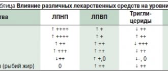
All married couples included in the study were examined in accordance with the order of the Ministry of Health of Russia No. 107n dated August 30, 2012 “On the procedure for using assisted reproductive technologies, contraindications and restrictions on their use” [3]. Before treatment with ART methods, all patients had the level of anthropogenic chemicals (ACS) in the blood determined by mass spectrometry. To assess the total level of ACV, a conditional scale, developed earlier, was developed ( Table 1
). The patients were divided into groups depending on the level of AChB: group 1 consisted of 72 patients with a high level of AChB (5 points or more), group 2 consisted of 72 patients with a low level of AChB. After dividing the patients into groups depending on the level of AChB (high/low), they were randomized to assess the effectiveness of antioxidant therapy.
Group 1, high level of AChB (n = 72):
- group 1a, antioxidant therapy+ (n = 36);
- group 1b, antioxidant therapy – (n = 36). Group 2, low level of AChB (n = 72);
- group 2a, antioxidant therapy+ (n = 36);
- group 2b, antioxidant therapy – (n = 36).
Antioxidant therapy in the experimental group was carried out for 2 months. before ART. For this purpose, coenzyme Q10 300 mg/day orally, eicosapentaenoic acid 300 mg/day orally, and docosahexaenoic acid 200 mg/day orally were used. All patients in the control group did not take antioxidant drugs for at least 6 months. before entering the ART cycle.
Ovarian stimulation was carried out according to a protocol with gonadotropin-releasing hormone antagonists; the dose of gonadotropins was selected individually. An ovulation trigger was introduced if there were follicles with a diameter of 17 mm or more in the ovaries. Human chorionic gonadotropin (HCG) 8000–10,000 IU or a gonadotropin-releasing hormone agonist 0.2 mg was used as an ovulation trigger. Support for the luteal phase and post-transfer period in all patients was carried out according to the standard protocol [7].
Fertilization of oocytes was carried out using intracytoplasmic sperm injection into the oocyte (ICSI). Cultivation and embryo transfer were carried out according to methods accepted in clinical practice. The proportion of mature oocytes was taken into account as the ratio of oocytes at the M2 stage to the total number of oocytes obtained. The fertilization rate was understood as the ratio of zygotes with normal fertilization to the total number of oocytes at the M2 stage. Blastocysts of excellent quality are embryos of classes 4AA, 5AA and 6AA according to the Gardner classification [8]. In all cases, selective embryo transfer was carried out in the native cycle.
14 days after transfer to the uterine cavity, the concentration of ß-CG in the patient’s blood serum was determined. When visualizing the heartbeat of the embryo after 5 weeks. After embryo transfer, clinical pregnancy was recorded. Statistical software package Statistica 10 (USA) was used for statistical analysis. Normally distributed data are presented as mean (standard deviation). Statistical analysis was carried out using the χ2 test to compare categorical variables, t test to compare means. Differences between statistical values were considered statistically significant at p < 0.05. The study was approved by the ethics commission of the National Medical Research Center of AGP named after. Academician V.I. Kulakov."
Introduction
Chronic cerebrovascular insufficiency is one of the most common syndromes in clinical neurology, incl.
among people of working age. The terminology of angiocerebral pathology is debatable; there is recognition of the validity of the concept “dyscirculatory encephalopathy”, which is a more general definition of the pathogenetic processes underlying brain damage of a vascular nature, incl. and ischemia, and microhemorrhage, and disorders of liquor dynamics, and neurogenic regulation of the microcirculatory bed. At the same time, in the existing classification of diseases of the circulatory system, the term “chronic cerebral ischemia” (CHI) remains stable, which is an “umbrella” diagnosis that unites an etiologically and pathogenetically diverse group of clinical conditions that develop as a result of vascular lesions of the brain (CM), not associated with strokes [1–3]. Etiological factors for the development of CCI can be divided into genetic (presence of the epsilon-4 allele of the APOE gene, cerebral autosomal dominant arteriopathy with subcortical infarctions and leukoencephalopathy - CADASIL), socio-demographic (age over 60 years, Mongoloid or Negroid race, male gender, low educational level level), general risk factors for the development of vascular diseases (arterial hypertension [AH], coronary heart disease, orthostatic hypotension, diabetes mellitus, hypercholesterolemia, hyperhomocysteinemia, obesity), others (smoking, alcoholism, sleep apnea) [4–6].
As the results of many studies show, it is cognitive impairment (CI) that represents the earliest and most objective manifestation of chronic vascular pathology of the brain; therefore, a rough estimate of the prevalence of CCI can be made based on studies of the prevalence of vascular CI conducted in Western countries. According to various researchers, cerebrovascular CIs are detected in 5–22% of elderly people [1]. At autopsy, certain vascular changes, most often of a microvascular nature, are found in about a third of elderly people. Thus, the overall prevalence of chronic cerebrovascular pathology may be about a third of elderly people.
The most important factors in the development of CCI are still vascular risk factors [7]: hypertension, dyslipidemia, diabetes mellitus, atrial fibrillation, overweight/obesity. There is an interrelation between these factors; their simultaneous presence significantly increases the risk of the occurrence and progression of CI [8]. Of course, coronary heart disease, accompanied by chronic heart failure, hemostatic disorders, venous discirculation, arterial hypotension, smoking, and alcohol abuse play an important role in the development and progression of CCI [8]. In this case, damage to both small arteries and large arterial trunks or a combination of both can form. With predominant damage to small-caliber arteries, either focal damage or diffuse damage to brain tissue develops in the form of the formation of microinfarctions and leukoaraiosis. Changes in large arteries lead to the formation of territorial and “watershed” cerebral infarctions. The listed risk factors present in patients contribute to the development of hemodynamic and hemorheological disorders [9].
The main pathogenetic mechanisms of cerebral ischemia constitute an “ischemic cascade”, including a decrease in cerebral blood flow, an increase in glutamate excitotoxicity, calcium accumulation and lactic acidosis, activation of intracellular enzymes, local and systemic proteolysis, the emergence and progression of antioxidant stress, the expression of early response genes with the development of depression of plastic protein and decrease in energy processes, long-term consequences of ischemia (local inflammatory reaction, microcirculatory disorders, damage to the blood-brain barrier).
The nature and severity of clinical disorders in CCI depend on the location, volume of the affected tissue and the number of lesions, while mnestic disorders, as a rule, do not dominate the clinical picture [10]. In such a situation, ranking the stages of CCI depending on the severity of pathomorphological changes in the brain becomes relevant.
To diagnose CCI, it is advisable to use the appropriate criteria [5]: 1) objectively identified neuropsychological and/or neurological symptoms; 2) signs of cerebrovascular disease, including risk factors and/or instrumentally confirmed signs of damage to cerebral vessels (for example, duplex scanning data), and/or GM substances (CT/MRI data); 3) the presence of a cause-and-effect relationship between vascular brain damage and the clinical picture of the disease; 4) absence of signs of other diseases that could explain the clinical picture.
Pathogenetic therapy for CCI should be aimed at optimizing cerebral blood flow and creating neurometabolic protection of the brain from ischemia and hypoxia. Treatment of patients with CCI primarily involves controlling vascular risk factors, preventing stroke and progression of chronic cerebrovascular pathology and, consequently, improving cognitive function. Prevention of stroke is based on correction of its risk factors (smoking, alcohol abuse, low physical activity, obesity), treatment of hypertension and diabetes mellitus [11]. Regular physical activity in the form of walking and therapeutic exercises is recommended, the intensity and duration of which depend on the functional state of the patient and the presence of concomitant diseases; Swimming can be helpful in many cases. It is recommended to include a large amount of fruits and vegetables with antioxidant properties in the diet [12].
Complex therapy for CCI includes the prescription of antioxidants, antiplatelet agents, drugs that optimize GM metabolism, and vasoactive drugs. Antidepressants are prescribed for severe asthenodepressive manifestations of the disease. Antiasthenic drugs are prescribed in a similar way.
An important component of the treatment of CCI is the administration of drugs with antioxidant activity. Currently, the following drugs of this series are used in clinical practice: Mexidol, Trollox, tocopherol acetate, tocopherol succinate, exiphon, tirilazad, meclofenoxate, atherovit, ebselen, thiotriazoline, emoxypine, cytoflavin, glutoxim [13].
Clinical studies [14] have revealed the promise of using ethylmethylhydroxypyridine succinate to alleviate the manifestations of asthenic syndrome, psychoemotional disorders and cochleovestibular disorders in patients with CCI. The drug helped reduce insulin resistance, hypertriglyceridemia, and hyperglycemia in patients with cerebrovascular diseases and metabolic syndrome [15].
Purpose of the study: to study the effectiveness and safety of therapy with the antioxidant drug ethylmethylhydroxypyridine succinate at a dose of 250 mg 3 times a day for 60 days for patients with CCI due to hypertension and atherosclerosis.
Objectives of the study: 1) to evaluate the effectiveness of ethylmethylhydroxypyridine succinate 250 mg as part of complex therapy for patients with CCI against the background of standard therapy for 60 days when administered orally compared with control; 2) evaluate the safety of ethylmethylhydroxypyridine succinate 250 mg as part of complex therapy for patients with CCI against the background of standard therapy for 60 days when administered orally compared with control; 3) to study the effect of a course of parenteral and oral administration of ethylmethylhydroxypyridine succinate 250 mg on the dynamics of neurological and neuropsychiatric manifestations of CCI in comparison with control.
Methods
The study included 40 patients aged 45–75 years who had a diagnosis of CCI verified by neuroimaging methods (CT or MRI of the brain), with signs of a stable course of the disease for at least a month before screening, in the presence of hypertension, atherosclerosis, and the absence of drug therapy with vasoactive drugs. nootropic, neurotrophic, neuroregenerative and antioxidant effects, incl. with the active ingredient ethylmethylhydroxypyridine succinate, 3 months before inclusion in the study. Exclusion criteria: age less than 45 and more than 75 years, presence of stroke 6 months before inclusion in the study, presence of myocardial infarction 6 months before inclusion in the study, poorly controlled hypertension with blood pressure numbers more than 200/100 mm Hg. , the presence of dementia, diabetes mellitus, congestive heart failure of functional class II or more, severe liver and kidney failure, cancer; taking vasoactive, nootropic, neurotrophic, neuroregenerative and antioxidant drugs 3 months before inclusion in the study, pregnancy and lactation, established hypersensitivity to ethylmethylhydroxypyridine succinate, alcohol abuse, participation in other clinical trials, the presence of other symptoms/diseases, in the opinion of the investigator, that could interfere patient's participation in the study or influence test results.
All patients included in the study, in addition to objective examination methods (blood pressure, heart rate, etc.), a detailed neurological examination, were assessed for the dynamics of subjective complaints of patients using the clinician's General Clinical Impression scale (Clinical Global Impression; CGI, W. Guy, 1976), Montreal Cognitive Rating Scale (MoCA, Z. Nasreddine, 2004), Subjective Asthenia Rating Scale (MFI 20, EM Smets et al., 1994), Hamilton Anxiety Rating Scale (HARS, M. Hamilton, 1959) and Hamilton Depression Scale, as well as assessment of motor activity in the elderly (Tinetti scale, M. Tinetti, 1986).
Patients included in the study were randomized into two groups using the envelope method. Patients of the 1st (main) group (20 people) were prescribed ethylmethylhydroxypyridine succinate orally at a dose of 250 mg 3 times a day for 60 days from the start of inclusion in the study; Patients of the 2nd (control) group underwent correction of existing risk factors (hypotensive, hypolipidemic, antiplatelet or anticoagulant therapy, correction of emotional status) in accordance with accepted clinical recommendations, guidelines for the management of hypertension, atherosclerosis, CCI.
All patients signed informed consent for inclusion in the study.
Statistical processing of its results was carried out using the application package Statistics 8.0. The degree of statistical significance of differences between unrelated groups was assessed using the nonparametric Mann–Whitney test. The degree of statistical significance of differences between related groups was assessed using the nonparametric Wilcoxon test. Differences were considered significant at the p<0.05 level.
Research results
The study was conducted on the basis of the Department of Nervous Diseases with a course of medical rehabilitation of the Krasnoyarsk State Medical University named after. Professor V.F. Voino-Yasenetsky from 04/01/2019 to 07/01/2019. The study underwent ethical review by the local ethics committee of Krasnoyarsk State Medical University.
When examining patients at the time of inclusion in the study, the following results were obtained: 40 patients were included, of which 29 (72.5%) were women and 11 (27.5%) men aged from 52 to 72 years, the median age in the first group was 59 ,1 [56; 71] years, in the second – 58.9 [57; 70]. The groups did not differ in gender and age composition, or the severity of vascular risk factors.
Patients of groups 1 and 2 complained of headache in 72 and 66% of cases, respectively, dizziness in 38 and 32%, unsteadiness in 12 and 16%, mood changes in 56 and 34%, decreased memory , concentration of attention – in 23 and 30% of cases.
Based on the assessment of the condition at the time of inclusion in the study using clinical scales, the data presented in Table 1 were obtained. 1.
As can be seen from the presented data, the groups were homogeneous in terms of the severity of complaints, as well as vestibulo-atactic, asthenic, cognitive, emotional-volitional disorders. The degree of severity of asthenia in both groups corresponded to the clinically expressed one, in patients of the main and control groups, emotional-volitional disorders corresponded to symptomatic anxiety, mild depression, the level of CI corresponded to moderate disorders in both groups, motor activity on the Tinetti scale corresponded at the time of inclusion in both groups to its moderate decrease.
Repeated examination of patients after 2 weeks of treatment revealed satisfactory tolerability of therapy; there were no cases of discontinuation of the test drug due to intolerance in the main group; 3 people in the main group and 1 in the control group developed a moderate headache that did not require additional therapy or changes in the treatment regimen . At the same time, in a number of patients, complaints decreased: for example, in the 2nd week of treatment, patients of the 1st and 2nd groups complained of headache in 68 and 65% of cases, respectively, of dizziness - in 28 and 33%, of unsteadiness - in 12 and 16%, for mood changes - in 30 and 24%, for decreased cognitive functions - in 24 and 30% of cases.
When re-testing the set of scales used, the indicators presented in table were obtained. 2.
Repeated assessment of the scales indicates a trend toward improvement in the Tinneti Balance Scale, the Hamilton Anxiety and Depression Scale, and the Clinical Global Impression Scale. Patient compliance in both groups was high, and continuation of therapy was recommended.
When re-evaluating the patients' condition at the end of the course of treatment (after 2 months of therapy), the following changes occurred: in groups 1 and 2, the number of patients complaining of headache decreased by 60 and 5%, respectively, and dizziness - by 50 and 15%, respectively, for unsteadiness - by 70 and 10%, mood changes - by 80 and 27%, CI - by 50 and 12%.
Using a set of validated scales, the data presented in table were obtained. 3.
As follows from the presented data, as a result of treatment with the drug ethylmethylhydroxypyridine succinate 250 mg in the main group, the severity of asthenia symptoms according to the subjective assessment scale MFI-20 significantly decreased from the initial severe asthenia with a median of 48 [47; 57] to normal values – 16 [10; 17] (p <0.0001, Wilcoxon test), while the antiasthenic effect of the drug appeared already in the early stages of treatment. Assessment of cognitive functions at the end of treatment showed an increase in mental performance and pace of task completion, while the Montreal Cognitive Function Assessment Scale indicators had positive dynamics in the main group of the disease with a median at inclusion in the study of 22 [20; 25], at the end of the treatment course – 24 [21; 26] (p=0.026, Wilcoxon test).
The regression of complaints of dizziness and unsteadiness at the end of treatment was accompanied by an increase in the assessment of motor activity of the elderly on the Tinetti scale from a moderate degree of impairment upon inclusion in the study (32 [23; 33]) to a mild degree at the end of the course of therapy: 37 [28; 38] (p<0.001, Wilcoxon test).
Evaluation of the examined patients of both groups on the general impression scale revealed an advantage in the main group of patients (p = 0.047, Mann-Whitney test) who were treated with the drug ethylmethylhydroxypyridine succinate 250 mg compared with patients who received treatment as part of the correction of risk factors in accordance with generally accepted standards for the management of hypertension and atherosclerosis.
Discussion
Chronic cerebrovascular diseases are conditions that doctors of various specialties most often encounter in everyday practice, so scientific research into this problem does not lose its relevance. A variety of pathological conditions underlying the development of CCI predetermine the formation of angioencephalopathy, which is manifested by various neuropsychiatric disorders, often identified in foreign literature as independent nosological forms, for example, multi-infarct leukoencephalopathy, Binswanger's disease, etc. Pathogenetic therapy of CCI should be aimed at optimizing cerebral blood flow and creation of neurometabolic protection of the brain from ischemia and hypoxia. That is why, along with the correction of vascular risk factors, antioxidant drugs are attracting the attention of researchers.
Ethylmethylhydroxypyridine succinate was studied and developed at the Research Institute of Pharmacology of the Russian Academy of Medical Sciences and the All-Union Scientific Center for the Safety of Biologically Active Substances [16]. The drug has a wide range of pharmacological effects, has antioxidant, antihypoxic, anxiolytic, antistress, antialcohol, anticonvulsant, neuroprotective, nootropic, antimnestic, neuropsychotropic, cardioprotective, antiatherosclerotic, antiplatelet, antiparkinsonian, vegetotropic effects. Clinical studies [17–19] have demonstrated significant therapeutic effects of the drug in the treatment of neurological, mental and cardiovascular diseases.
Thus, ethylmethylhydroxypyridine succinate has shown effectiveness in the treatment of neurotic and neurosis-like disorders, various conditions of alcoholism, incl. withdrawal syndrome, as well as acute and chronic disorders of cerebral circulation (stroke, discirculatory encephalopathy, vegetative-vascular dystonia), dysfunction of the brain during aging and atherosclerosis.
A significant advantage of the drug is its wide range of pharmacological effects, low toxicity, virtual absence of side effects (typical, for example, for some “standard” neuropsychotropic drugs), sedative, muscle relaxant, stimulating and euphoric effects [18].
Conclusion
This paper presents the results of the use of ethylmethylhydroxypyridine succinate at a dosage of 250 mg 3 times a day for 2 months in patients with CCI caused by hypertension and atherosclerosis. The results of the use of the drug indicate the effectiveness of treating the main neurological and neuropsychiatric symptoms of CCI, such as dizziness, ataxia, CI, anxiety and asthenia. As a result of treatment with ethylmethylhydroxypyridine succinate 250 mg in the main group, there was a significant decrease in the severity of asthenia symptoms according to the subjective MFI-20 assessment scale, while the anti-asthenic effect appeared already in the early stages of treatment. Assessment of cognitive functions at the end of treatment showed an increase in mental performance and the pace of completing tasks according to the Montreal Cognitive Function Assessment Scale; regression of complaints of dizziness and unsteadiness at the end of treatment was accompanied by an increase in the assessment of motor activity in the elderly according to the Tinetti scale. Over the course of 2 months, the drug demonstrated safety when used at the indicated dosage. The identified side effects were short-lived and minor.
results
The study included 144 patients who were divided into groups depending on the level of AChB in the body and the prescription of antioxidant therapy. Clinical characteristics of the patients included in the study are presented in Table. 2
. When comparing clinical and anamnestic characteristics, no differences were found between the subgroups.
When assessing the parameters of ovarian stimulation (duration of stimulation, total and daily dose of gonadotropins, frequency of use of various drugs), no differences were also found.
Then we assessed the parameters of folliculo-, oo- and early embryogenesis in the comparison groups.
In the group of patients who had low levels of pollutants and received antioxidant therapy, a decrease in the number of follicles and oocytes was observed. The median number of follicles was 8 in group 2a compared with 10 in group 1a, 11 in group 1b and 12 in group 2b (p = 0.0645). The median number of oocytes was 7 in group 2a compared with 9 in group 1a, 10 in group 1b and 9 in group 2b (p = 0.0276). In the group of patients who had a high level of pollutants and received antioxidant therapy, a decrease in the frequency of fertilization was observed: the median frequency of fertilization in group 1a was 90.9 compared to 100 in groups 1b, 2a and 2b (p = 0.0973); table 3
.
When assessing the clinical outcomes of ART cycles, an increase in the incidence of clinical pregnancy and live births was observed in groups using antioxidant therapy ( Table 4
). When comparing within groups (groups 1a/1b and 2a/2b), these differences did not reach statistical significance.
Table 1. Scale for assessing the total level of pollutants in the body
| Pollutant | ≥50%, 1 point | ≥75%, 2 points | ≥97.5%, 3 points |
| Lead, µg/l | 9,85 | 13,59 | 35,36 |
| Cadmium, µg/l | 0,32 | 0,45 | 1,45 |
| Mercury, µg/l | 0,81 | 1,44 | 4,50 |
| Bisphenol A, ng/ml | 0,52 | 0,66 | 12,65 |
Table 2. Clinical characteristics of patients in comparison groups
| Group 1a, n=36 | Group 1b, n=36 | Group 2a, n=36 | Group 2b, n=36 | R** | |
| Age, years* | 32 (28–34) | 32 (29–35) | 31 (28–33) | 30 (28–33) | 0,3896 |
| BMI, kg/m2* | 21,7 (19,8–23,0) | 21,6 (20,4–23,8) | 21,2 (20,0–23,5) | 21,5 (20,6–22,5) | 0,8618 |
| Number of pregnancies in history | 0 (0–1) | 1 (0–1) | 0 (0–1) | 0 (0–1) | 0,8616 |
| Primary infertility, n (%) | 18 (50,0) | 18 (50,0) | 21 (58,3) | 19 (52,8) | 0,8821 |
| Duration of infertility, years* | 4 (2–6) | 4 (3–5) | 4 (2–5) | 4 (2,5–6) | 0,7340 |
| Number of ART cycles in history | 0 (0–1) | 0 (0–1) | 0 | 0 (0–1) | 0,0591 |
| AMG, ng/ml* | 3,1 (1,4–5,5) | 2,8 (1,5–5,8) | 2,9 (1,9–6,3) | 3,9 (2,3–6,9) | 0,2624 |
| FSH, mIU/ml* | 6,1 (4,7–7,6) | 6,7 (5,2–7,5) | 6,6 (5,3–8,1) | 6,6 (5,4–7,6) | 0,6920 |
*Data are presented as median (interquartile range). **Kruskal–Wallis test for comparison of continuous data, χ2 for comparison of categorical data
Table 3. Parameters of the embryological stage in patients in comparison groups
| Group 1a, n = 36 | Group 1b, n = 36 | Group 2a, n = 36 | Group 2b, n = 36 | R** | |
| Number of follicles* | 10 (6–15) | 11 (7–14) | 8 (6–11) | 12 (8–14) | 0,0645 |
| Number of oocytes* | 9 (5–14) | 10 (6–12) | 7 (5–10) | 9 (8–13) | 0,0276 |
| Number of oocytes, metaphase II* | 8 (4–10) | 7 (5–11) | 5 (3–8) | 6 (5–10) | 0,2933 |
| Proportion of mature oocytes* | 0,80 (0,67–0,93) | 0,83 (0,71–1,0) | 0,83 (0,71–1,0) | 0,76 (0,55–0,95) | 0,2415 |
| Fertilization frequency* | 90,9 (77,5–100) | 100 (80,9–100) | 100 (83,8–100) | 100 (95–100) | 0,0973 |
| Number of blastocysts* | 2 (1–5) | 3 (1–4) | 3 (1–4) | 3 (1–4) | 0,9527 |
| Number of blastocysts of excellent quality* | 2 (1–4) | 2 (1–4) | 2 (1–4) | 2 (1–4) | 0,8890 |
| Blastulation rate* | 0,35 (0,25–0,50) | 0,41 (0,18–0,63) | 0,50 (0,35–0,71) | 0,50 (0,17–0,67) | 0,0903 |
| Availability of embryos suitable for cryopreservation | 22 (61,1%) | 24 (66,7%) | 26 (72,2%) | 22 (61,1%) | 0,7181 |
*Data are presented as median (interquartile range). **Kruskal–Wallis test for comparison of continuous data, χ2 for comparison of categorical data
Table 4. Results of ART cycles in comparison groups
| Group 1a, n = 36 | Group 1b, n = 36 | OR 1a/1b (95% CI) | Group 2a, n = 36 | Group 2b, n = 36 | OR 2a/2b (95% CI) | OR 1/2 (95% CI) | |
| Pregnancy Rate | 18 (50,0%) | 11 (30,6%) | 2,27 (0,78; 6,70) | 24 (66,7%) | 16 (44,4%) | 2,50 (0,87; 7,27) | 2,33 (1,14; 4,81) |
| Birth rate | 12 (33,3%) | 9 (25,0%) | 1,50 (0,48; 4,79) | 19 (52,8%) | 14 (38,9%) | 1,76 (0,62; 4,98) | 1,61 (0,77; 3,37) |
| Ʃ birth rate | 17 (47,2%) | 12 (33,3%) | 1,79 (0,62; 5,19) | 24 (66,7%) | 17 (47,2%) | 2,24 (0,78; 6,48) | 1,96 (0,96; 4,02) |
Drawing. Results of ART cycles in comparison groups
When comparing groups 1 and 2, a statistically significant increase in the pregnancy rate in group 1 was obtained (OR 2.33, 95% CI 1.14; 4.81), however, the differences in assessing the frequency of births and the cumulative frequency of births also did not reach statistical significance. The obtained data are presented in table. 4
and in
Fig
.
The number of patients treated (NNT) was calculated. In the subgroup of patients with high levels of ACV (group 1), the absolute risk reduction (ARR) was calculated using the formula 18/36 – 11/36 = 0.5 – 0.306 = 0.194. NPL was calculated using the formula 1/0.194 = 5.2 = 5.
In the subgroup of patients with low levels of AChB (group 2), ASR was calculated using the formula 24/12 – 16/20 = 0.667 – 0.444 = 0.223. NLP was calculated using the formula 1/0.223 = 4.5 = 5. In the general group of patients, ASR was calculated using the formula 42/72 – 27/72 = 0.583 – 0.375 = 0.208. NPL was calculated using the formula 1/0.208 = 4.8 = 5.
Discussion
Many patients undergoing fertility treatment take various vitamins and antioxidant medications in hopes of improving their fertility. For most patients, preparation for ART programs is associated with great emotional stress. Conducting studies to evaluate the effectiveness of antioxidant drugs is important because patients must be provided with objective research data that will allow them to make decisions about taking these drugs. The complexity of studying this problem is due to several factors. First, it is difficult to account for dietary antioxidants, and dietary and lifestyle factors may also influence reproductive health. Secondly, most of the oral antioxidants are registered as dietary supplements; they are sold not only through pharmacy chains, but also in supermarkets/sports clubs, which makes them difficult to account for. In addition, non-medicinal products are not subject to comprehensive pharmacovigilance, therefore control of the dosage of active components and the quality of the active substance remains the task of the manufacturer [9, 10]. Third, different studies have examined different doses and drugs, and use different parameters as endpoints (sperm quality, embryological characteristics, results of ART cycles), which, in turn, may depend on the presence of multiple risk factors.
Previous studies have shown the negative impact of ACV on the quality of gametes and embryos in ART cycles [6, 11]. The main mechanism of influence of environmental factors on reproductive health is the induction of oxidative stress, and the main mechanism of antioxidants is the neutralization of free radicals, as well as the activation of the synthesis of antioxidant enzymes [12]. The choice of drugs for antioxidant therapy was based on literature data [13–15].
Clinical and anamnestic characteristics did not differ between groups. When assessing the embryological stage, a trend toward a decrease in the total number of oocytes in the experimental group was observed, but no differences were recorded in parameters reflecting the quality of oocytes (number of mature oocytes, proportion of mature oocytes in the overall cohort). There was also a tendency towards a decrease in the frequency of fertilization in the group of patients with high levels of AChB who received antioxidant therapy (group 1a). Similarly, the blastulation rate was slightly higher in the group of patients with low AChB levels, although the differences did not reach statistical significance. Literature data on the effect of antioxidant therapy on embryological parameters of ART cycles are contradictory. Some studies have not shown the effect of drugs on the quality of oocytes and embryos [12, 16]. However, other studies have demonstrated an increase in embryo quality when administered a “Mediterranean diet” [17] or oral antioxidants [18]. The authors of the above studies suggest a positive effect of antioxidants on metabolic processes in the ovaries, but emphasize the need for further research in this area.
When assessing the clinical results of ART cycles, a trend towards an increase in the incidence of clinical pregnancy, birth rate and cumulative birth rate in subgroups using antioxidant therapy was noted. It is interesting to note that these differences were observed in both high AChB and low AChB subgroups, suggesting the involvement of additional mechanisms of action of antioxidant drugs.
The number of patients treated was 5 both for the overall group of patients and for subgroups depending on the level of ACV.
The results obtained allow us to recommend the use of antioxidant therapy to prepare patients for ART programs.
The strengths of this study were the randomized design, the use of a scale to assess the level of AChB in the body, the use of one ART protocol for all patients, and the assessment of all clinical outcomes, including the cumulative incidence of live births.
The weakness was the small sample size, which did not allow us to obtain statistically significant differences in the effectiveness of ART in patients with different levels of ACV.
Pathophysiology of oxidative stress
The basic mechanisms of pathology in any critical condition, including surgical and traumatic aggression, are free radical processes and changes in the properties of cell biomembranes. The main pathological role of free radicals is that they actively interact with molecules that form neuronal and intracellular membranes. The viscosity of the membranes increases, their plasticity and functional state are lost. Along with this, genes responsible for programmed cell death - apoptosis - are activated. There is a direct relationship between the accumulation of lipid peroxidation products and the severity of damage to nerve cells and other tissues.
Since the formation of tissue hypoxia, lipid peroxidation, and mitochondrial dysfunction are recognized as the trigger for the development of a typical pathological process, the use of antihypoxants and antioxidants is pathogenetically justified in surgical and traumatic aggression, inflammation and acute pain.
Restoring blood flow in previously ischemic tissues also poses a certain danger. Reperfusion causes a multiple increase in the partial pressure of oxygen with a further increase in free radical processes. In this case, the endothelium of the capillaries is damaged, the anticoagulant activity of which is transformed into procoagulant activity. Due to increasing adhesion, leukocytes and platelets clog the capillaries. This process is aggravated by an increase in the rigidity of red blood cells, which sharply increases the disruption of oxygenation of tissues, especially the brain. The processes of blood fibrinolysis are inhibited, the area of cerebral infarction expands, and cerebral edema increases.
The main pathological processes initiated by excessive activation of LPO
I. Cellular-tissue level:
- ischemia;
- hypoxia;
- membranopathy:
– disruption of the permeability of the cell membrane and membranes of cell organelles;
– excessive accumulation of free radicals inside the cell;
– release of lysosomal enzymes into the cell;
– accumulation of Ca++ ions inside the cell;
- apaptosis and cell necrosis;
- violation of cellular reception;
- energy and metabolic disorders.
II.
Organs and systems:
- functional disorders;
- organic pathology.
Of course, the body has an endogenous antioxidant system (AOS), but at critical levels of hypoxia and LPO it is untenable and it is necessary to introduce antioxidants from the outside.
conclusions
Detoxification therapy in preparation for assisted reproduction cycles helps to increase the effectiveness of ART cycles.
The results obtained allow us to recommend the use of antioxidant therapy to prepare patients for ART programs.
List of literature / References
- Thoma ME, McLain AC, Louis JF, King RB, Trumble AC, Sundaram R., Buck Louis GM Prevalence of infertility in the United States as estimated by the current duration approach and a traditional constructed approach. Fertil Steril. 2013;99(5):1324-1331.e1. https://doi.org/10.1016/j.fertnstert.2012.11.037.
- Chandra A., Copen CE, Stephen EH Infertility and impaired fecundity in the United States, 1982-2010: data from the National Survey of Family Growth. Natl Health Stat Report. 2013;(67):1-18. Available at: https://pubmed.ncbi.nlm.nih.gov/24988820
- Pizzol D., Foresta C., Garolla A., Demurtas J., Trott M., Bertoldo A., Smith L. Pollutants and sperm quality: a systematic review and meta-analysis. Environ Sci Pollut Res Int. 2021;28(4):4095-4103. https://doi.org/10.1007/s11356-020-11589-z.
- Fichman V., Costa RSSD, Miglioli TC, Marinheiro LPF Association of obesity and anovulatory infertility. Einstein (Sao Paulo). 2020;18:eAO5150. https://doi.org/10.31744/einstein_journal/2020AO5150.
- Carvalho LVB, Hacon SS, Vega CM, Vieira JA, Larentis AL, Mattos RCOC et al. Oxidative Stress Levels Induced by Mercury Exposure in Amazon Juvenile Populations in Brazil. Int J Environ Res Public Health. 2019;16(15):2682. https://doi.org/10.3390/ijerph16152682.
- Syrkasheva A.G., Frankevich V.E., Dolgushina N.V. Association between the level of heavy metals in the body of women with infertility and the outcomes of assisted reproductive technology programs. Obstetrics and gynecology. 2020;(11):124-130. https://doi.org/10.18565/aig.2020.11.124-130.
- Syrkasheva A.G., Dolgushina N.V., Makarova N.P., Kovalskaya E.V., Agarsheva M.A. Outcomes of assisted reproductive technology programs in patients with oocyte dysmorphisms. Obstetrics and gynecology. 2015;(7):56-62. Access mode: https://aig-journal.ru/articles/Ishody-programm-vspomogatelnyh-reproduktivnyh-tehnologii-upacientok-s-dismorfizmami-oocitov.html.
- Gardner D., Schoolcraft WB Culture and transfer of human blastocysts. Curr Opin Obs Gynecol. 1999;11(3):307-311. https://doi.org/10.1097/00001703-199906000-00013.
- Kalugina A.S., Bespalova A.N., Kovaleva I.V., Bakleicheva M.O. The effectiveness of using a vitamin-mineral complex during pregnancy. Pharmateka. 2019;26(6):74-78. https://doi.org/10.18565/pharmateca.2019.6.73-78.
- Brough L., Rees GA, Crawford MA, Morton RH, Dorman EK Effect of multiple-micronutrient supplementation on maternal nutrient status, infant birth weight and gestational age at birth in a low-income, multiethnic population. Br J Nutr. 2010;104(3):437-445. https://doi.org/10.1017/S0007114510000747.
- Syrkasheva A.G., Kindysheva S.V., Starodubtseva N.L., Frankevich V.E., Dolgushina N.V. The influence of bisphenol A on the outcomes of assisted reproductive technology programs in patients with infertility. Gynecology. 2021;23(2):161-166. Available at: https://omnidoctor.ru/library/izdaniya-dlya-vrachey/ginekologiya/gn2021/gn2021_23_2/vliyaniebisfenola-a-na-iskhody-programm-vspomogatelnykh-reproduktivnykhtekhnologiy-u-patsientov
- Zarezadeh R., Mehdizadeh A., Leroy JLMR, Nouri M., Fayezi S., Darabi M. Action mechanisms of n-3 polyunsaturated fatty acids on the oocyte maturation and developmental competence: Potential advantages and disadvantages. J Cell Physiol. 2019;234(2):1016-1029. https://doi.org/10.1002/jcp.27101.
- Ohsawa I., Ishikawa M., Takahashi K., Watanabe M., Nishimaki K., Yamagata K. et al. Hydrogen acts as a therapeutic antioxidant by selectively reducing cytotoxic oxygen radicals. Nat Med. 2007;13(6):688-694. https://doi.org/10.1038/nm1577.
- Falsig A.-ML, Gleerup CS, Knudsen UB The influence of omega-3 fatty acids on semen quality markers: a systematic PRISMA review. Andrology. 2019;7(6):794-803. https://doi.org/10.1111/andr.12649.
- Chiu Y.-H., Karmon A.E., Gaskins A.J., Arvizu M., Williams P.L., Souter I. et al. Serum omega-3 fatty acids and treatment outcomes among women undergoing assisted reproduction. Hum Reprod. 2018;33(1):156-165. https://doi.org/10.1093/humrep/dex335.
- Lass A., Belluzzi A. Omega-3 polyunsaturated fatty acids and IVF treatment. Reprod Biomed Online. 2019;38(1):95-99. https://doi.org/10.1016/j.rbmo.2018.10.008.
- Kermack AJ, Lowen P, Wellstead SJ, Fisk HL, Montag M, Cheong Y et al. Effect of a 6-week “Mediterranean” dietary intervention on in vitro human embryo development: the Preconception Dietary Supplements in Assisted Reproduction double-blinded randomized controlled trial. Fertil Steril. 2020;113(2):260-269. https://doi.org/10.1016/j.fertnstert.2019.09.041.
- Xu Y., Nisenblat V., Lu C., Li R., Qiao J., Zhen X., Wang S. Pretreatment with coenzyme Q10 improves ovarian response and embryo quality in lowprognosis young women with decreased ovarian reserve: a randomized controlled trial . Reprod Biol Endocrinol. 2018;6(1):29. https://doi.org/10.1186/s12958-018-0343-0.

![Rice. 1. ACCOMPLISH study: the effect of the combination of ACE inhibitor* amlodipine and ACE inhibitor hydrochlorothiazide on the risk of primary endpoint events (CVE) [14]](https://expert35.ru/wp-content/uploads/ris-1-issledovanie-accomplish-vliyanie-kombinacii-iapf-amlodipin-i-iapf-330x140.jpg)