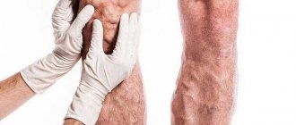
The veins of the lower extremities ensure uninterrupted outflow of blood, participate in the regulation of blood pressure, and transport hormones, enzymes, amino acids and other organic substances. The vascular system of the legs is the most vulnerable because it is far from the heart, but at the same time experiences constant physical stress. Impaired blood flow and depletion of blood vessels leads to their stretching and rupture.
Telangiectasia - dilation and rupture of small vessels (capillaries, venules and arterioles) is most often expressed only in external cosmetic defects. If a vein in the leg bursts, it is accompanied by pain and hemorrhage. In severe cases, perforation of the vascular wall is accompanied by severe bleeding, requiring emergency medical attention.
Differences between vessels
Venous and arterial vessels should not be confused. In the body, these elastic tubes perform various functions and differ in structure, location and number. It’s a vein that can burst in your leg, not an artery, and here’s why:
- The artery has thick walls with many smooth muscle fibers and can withstand the powerful pressure of the blood flow. Venous walls are several times thinner.
- The veins along their entire length are equipped with valves that prevent reverse blood flow. Arterial valves are present only at the exit of blood vessels from the heart.
- The arteries are located deep under the skin, protruding only in a few areas of the body where the pulse can be felt (neck, temples, wrist). The venous network is located superficially.
- Arterial vessels carry oxygenated blood from the heart to the organs. Venous blood, filled with carbon dioxide, moves in the opposite direction.
When an artery ruptures, blood is ejected in a fountain - this condition is life-threatening. Venous bleeding is characterized by the uniform flow of a smaller volume of blood into the subcutaneous tissue (internal) or outside the body (external).
What are the dangers of injuries to large vessels?
When a large artery and vein are simultaneously damaged, a serious complication such as an arteriovenous fistula
. This, in turn, causes the appearance of a false arteriovenous aneurysm. Its main danger is that it often becomes inflamed with the formation of pus. The consequences of opening such phlegmons are very serious. Sometimes vascular injuries are diagnosed only after the appearance of such aneurysms.
Arteriovenous fistulas are found quite often, but the most dangerous of them is a fistula in the neck area. It can cause heart failure. If the fistula is not recognized in time, gangrene may develop.
Briefly about the structure of venous walls
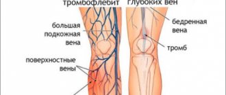
The venous system of the lower extremities consists of superficial and deep vessels connected by a network of perforating ducts. The walls of the veins consist of three layers, each of which is endowed with certain functional responsibilities:
- Endothelium (intima) or inner layer. Protects against the effects of substances carried through the bloodstream.
- Media or middle layer. Consists mainly of elastic fibers responsible for tension/relaxation of the wall and vascular tone.
- Adventitia or outer layer. Contains collagen fibers that connect the vessel with muscle tissue and microscopic vessels that supply oxygen to the wall itself. Responsible for protection against perforation.
When the walls are dysfunctional, due to their exhaustion or extreme tension, the veins rupture.
Reference! Angiopathy of the lower extremities is a systemic lesion of blood vessels of all sizes. The pathology is corrected by an angiosurgeon. Varicose veins of the legs - expansion and increase in the lumen of the veins with the formation of nodes. Conservative or surgical treatment is carried out by a phlebologist.
During pregnancy
A separate case is the period of pregnancy in women. The body of the expectant mother experiences enormous stress: organs and systems are rebuilt, hormonal levels change. It is during this period that varicose veins are most often first detected in women.
Related factors play a huge role here:
- hemorrhagic diathesis, in which the smallest vessels burst,
- hereditary thrombophilia is a phenomenon accompanied by the appearance of hematomas for no apparent reason,
- some types of anemia can cause vascular damage,
- liver failure,
- taking certain medications.
Helpful information! Women carrying children must adhere to a diet, eat well and wear special underwear. These simple measures will help strengthen blood vessels and prevent further development of varicose veins.
Causes of vein rupture
The key reason why leg veins burst is varicose veins. Deformation of the vascular walls occurs due to the destruction of the valves. Blood begins to move not only up (towards the heart), but also down. This creates excess pressure on the walls.
The veins no longer cope with the load, expand, lengthen, bend, taking on tortuous shapes. Trophism (the process of cellular nutrition) and innervation (supply of nerves) of the lower extremities are disrupted. There are three stages of the disease:
- compensated - telangiectasia, single dilated vessels on one or both legs, without obvious symptoms;
- subcompensated – pronounced external changes in the veins, the appearance of nodes, swelling of the limbs, pain, cramps at night, eczema;
- decompensated - symptoms of subcompensation intensify, nodes progress, pre-ulcers and trophic ulcers form.
The risk of vein destruction exists from the first stage and only increases in the future. In the presence of varicose veins, triggers for vascular destruction are:
- nicotine addiction;
- alcohol abuse;
- excess weight;
- hormonal changes, including pregnancy;
- sedentary lifestyle (physical inactivity) or excessive physical activity;
- deficiency of the vitamin and mineral component of the diet;
- tissue damage caused by high or low temperatures (burns or frostbite);
- chronic diseases of the cardiovascular and endocrine systems.
An age-related decrease in the elasticity of blood vessels is associated with general wear and tear (aging) of the body. The development of internal venous bleeding can be triggered by taking medications that affect coagulation processes (blood clotting) or impaired peripheral circulation.
In the ankle area, perforation can occur due to constant wearing of narrow, compressive shoes. Open (external) venous bleeding occurs due to mechanical damage to the legs (deep wounds, animal bites, open fractures, etc.), with a violation of the integrity of the skin.
You need to perceive the situation correctly
It is known that before taking on a solution to a problem, it is necessary to understand it. In most cases, the culprit for the appearance of bruises and spider veins is varicose veins. For a long time it can proceed hidden and not cause much discomfort. A person begins to worry only when bruises appear on his legs. This sign means that the vessel has burst in this particular place. Vascular injury can occur anywhere in the lower extremity.
Important point! The regular appearance of hematomas (bruises) should be a serious argument for visiting a medical facility. You especially shouldn’t put off visiting a doctor when such symptoms are accompanied by pain and swelling in the affected area.
Signs of perforation
The main and clearly visible sign of a burst vein in the leg with hemorrhage into the subcutaneous layer is the appearance of a hematoma (accumulation of liquid, thickened or coagulated blood in the subcutaneous fat). The size and color of the resulting bruise can indicate the extent of the internal damage.
The accumulation of blood at the site of the vein rupture forms edema. A swelling of the tissues forms, which feels painful when pressed. With cracks in the veins or perforation of venules and capillaries, there are no significant somatic symptoms. More significant damage is accompanied by:
- pain in the limb (at the moment of the rupture - acute);
- paresthesia (sensitivity disorder in the legs, burning sensation, crawling “goosebumps” in the damaged area);
- paleness of the skin and mucous membranes;
- loss of strength (weakness) and dizziness;
- lack of air;
- polymorphic opacities in the field of vision (“flying spots before the eyes”);
- discomfort in the epigastric region (nausea).
Excessive internal bleeding can cause short-term fainting (syncope). External venous bleeding with damage to the skin is characterized by a slow flow of blood that has a specific dark cherry color, pain, severe nausea (vomiting may occur), and severe pallor of the skin. A person feels cold in the affected limb and chills throughout the body. Loss of consciousness is possible from painful shock (in especially impressionable people and from the sight of blood).
Clinical picture and diagnosis of blood vessel damage
Of course, one of the first symptoms of traumatic damage to blood vessels is the occurrence of characteristic bleeding
, however, it can only be seen with open wounds. Any intense bleeding should alert a specialist.
A simple palpation of the pulse slightly to the side of the wounded area can help make a correct diagnosis. However, the method is not 100% reliable. If a large arterial trunk is injured, a more accurate symptom will be the appearance of a swelling that quickly grows and pulsates. You can literally try to listen to such a wound, because such injuries produce a very noticeable blowing noise, sometimes compared to the purring of a cat.
In addition, you should pay attention to the skin at some distance from the wound: their pallor may also indicate damage to large vessels.
The most unambiguous sign that indicates major damage to the arteries and veins is gangrene of the limb
.
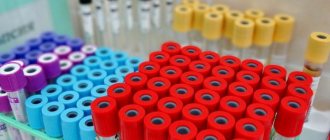
An accurate diagnosis is made after a vasographic X-ray contrast study. Capillaroscopy and thermography may be informative. Blood tests help in diagnosis, as well as a non-invasive research method such as Doppler sonography.
Treatment of internal venous hemorrhage
Primary treatment for mild to moderate tears is to cool the area of the leg. Under the influence of cold, the blood vessels narrow and the bleeding stops. Pharmacies sell special hypothermic packages for providing first aid for bruises and internal hemorrhages.
Hypothermic first aid package “Snowball”. Package composition: - ammonium nitrate, g 30+-3 - water, g 25+-5
At home, ice wrapped in cotton cloth or any frozen product from the freezer wrapped in cloth will be suitable for first aid. After applying the compress, the limb must be placed on a pillow or cushion to ensure blood flow in the desired direction.
In the future, a bruise caused by a burst vein is treated with drugs for external use that have anti-inflammatory, blood-thinning, anti-edematous, and angioprotective effects. Examples of gels and ointments:
- Traumeel;
- Bruise OFF;
- Troxevasin;
- Badyaga 911;
- Lyoton;
- Express Bruise;
- Heparin ointment;
- Viprosal.
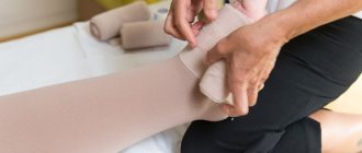
In case of a large-scale hematoma, it is necessary to apply a compressive bandage below the damaged area, carry out identical manipulations to cool the damaged area and seek medical help. For extensive internal bleeding and complicated hematomas, the following is practiced:
- puncture (pumping out) of biological fluid using a needle with a large lumen;
- surgical opening with a scalpel, followed by the application of self-absorbing sutures to the damaged vein.
The autopsy procedure is carried out under the influence of local anesthetics.
How to determine the nature of the lesion
Before planning treatment measures, it is necessary to accurately determine the type of hemorrhage. There are several simple tests that can quickly identify the cause of the damage.
- Pinch method. In the subclavian area, you need to use your fingers to gather a small area of skin into a fold, then lightly squeeze it and turn it. In the absence of vascular pathologies, a bruise does not form at the site of manipulation. If after the test a hematoma remains, it means the vessels are fragile.
- Hammer method. If after lightly tapping the chest with a percussion hammer a bruise appears, it can be judged that the blood vessels at the site of impact have burst.
- Tourniquet method. Testing is carried out using a rubber band or a tonometer cuff. The device must be placed on the middle of the shoulder and tightened. High fragility of blood vessels will be indicated by traces of pinpoint hemorrhages that appear after a five-minute test.
Conclusion. If the result of all three tests is positive, a person must undergo a full range of diagnostic procedures, because at any moment his capillaries or larger vessels may burst.
Important! The frequent appearance of petechiae, purpura, and bruises on the body should be regarded as a serious problem.
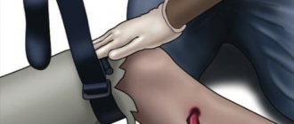
First aid for open injury
What to do if the vein and tissue are damaged, and blood continuously flows out? First of all, you should call an ambulance. Extensive venous bleeding without timely medical intervention threatens the loss of a large volume of blood and blockage of the blood vessel by air bubbles (air embolism).
Before the brigade arrives, the victim needs to receive emergency assistance. First of all, you should identify the damaged area and try to stop the bleeding. To do this, use a tourniquet (if not available, replace it with a belt, belt, etc.) and dressing material (medical special bags, gauze and bandage, any clean cloth).
Algorithm of actions:
- Lift the limb with the burst vein up and, placing available means under it, fix it in this position.
- Using a dressing material, tightly (important!) bandage the wound. Below the damaged area, tighten the bandage tightly to block the natural circulation of venous blood from bottom to top.
- A compressive tourniquet (belt) is applied a few centimeters below the bleeding area over clothing. If the limb is exposed, any fabric must be placed under the tourniquet. Be sure to record the time of the procedure on paper. You can clamp a limb for no more than one and a half hours. Otherwise, tissue necrosis may begin. Place paper with the time recorded under the tourniquet.
The use of a tourniquet is mandatory for arterial bleeding. If a vein is damaged, it is advisable to use a tourniquet when the bleeding does not stop after applying a bandage.
Characteristic symptoms
When a vessel is damaged, a hematoma forms almost immediately. A bruise can range in size from a few millimeters to tens of centimeters. In other cases, the spots occupy an impressive area, sometimes they merge, forming one huge hematoma. The formation can persist for several weeks, gradually changing color, and then disappearing without a trace.
Based on the type of hemorrhage, you can determine which vessel’s integrity is compromised:
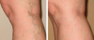
- Telangiectasia is a network of dilated small capillaries. Such vessels usually do not burst. This phenomenon is in the nature of a pronounced cosmetic defect. Most often, telangiectasias appear against the background of existing varicose veins.
- Purpura is the accumulation of some blood under the skin. The formation has the appearance of a smooth, shiny spot.
- Ecchymosis is a large bruise caused by trauma.
- Petechiae is a pattern in the form of pinpoint hemorrhages, which is formed by burst capillaries on the surface of the skin.
Important point! If the hemorrhage is not the result of an injury, you should immediately visit a doctor.
Vascular surgery of the lower extremities
After providing first aid for a burst vein, the next step should be surgery. The purpose of the intervention is to remove the damaged part of the vessel. Depending on the scale of the perforation and the general condition of the vessels, various methods of vascular surgery can be used:
- elos-coagulation – laser treatment of varicose veins using elos technology;
- sclerotherapy and its varieties (compression sclerotherapy, echosclerotherapy, microfoam sclerotherapy) - the introduction of a special drug into the vessels, leading to scarring of the vascular walls;
- miniphlebectomy is a minimally invasive technique for removing part of a vessel through a small puncture;
- Phlebectomy – surgical excision of a section of a damaged vein.
- laser coagulation – sealing of blood vessels with a laser beam.
The newest method used in phlebology is radiofrequency ablation - the adhesion of venous walls under the influence of radio wave radiation.
Diet
Proper nutrition is one of the important components of comprehensive treatment. To keep the blood vessels strong and not burst, you need to adjust the diet according to the diagram below.
What to give up
What foods are recommended to eat? Fatty meats, fast food, butter. Vegetable oils, nuts, lean fish, dietary meat. Alcohol, sweet desserts, smoked meats, chocolate, seasonings, spicy and salty dishes, marinades.
Fresh greens, fruits in unlimited quantities, garlic, cabbage, berries, any fruit and berry drinks, rosehip tea, lactic acid products.
Drug treatment
After vein perforation, the patient is prescribed a number of medications necessary to restore and maintain normal blood flow, eliminate formed blood clots, increase the tone of the vascular walls, prevent and eliminate the inflammatory process:
- venotonics for oral and external use - Venarus, Troxevasin, Detralex, etc.;
- anticoagulants – Thrombo Ass, Curantil, Fraxiparine;
- vitamins – vitamin complexes with a high content of ascorbic acid;
- thrombolytics (fibrinolytics) – Actylase, Retaplase, etc.;
- non-steroidal anti-inflammatory drugs (NSAIDs) - Ibuprofen, Diclofenac, Ketanov.
The doctor determines the dosage regimen and dosage individually.
Prevention
Varicose veins of the lower extremities must be treated from the first manifestations. To prevent the disease from passing into the decompensated stage, rupture of veins, and the occurrence of intermittent claudication, it is necessary to use preventive measures:
- wearing compression garments (stockings, tights, knee socks);
- refusal of nicotine and alcohol;
- regular classes of special therapeutic exercises for varicose veins;
- eating foods rich in vitamin C;
- daily contrast dousing of legs.
If you have problems with your veins, you should be regularly examined by a phlebologist.
How to prevent varicose veins
Basic questions about spider veins
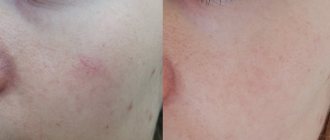
- Is it painful to remove spider veins on the legs?
This question worries all women who decide to get rid of spider veins on the skin. The answer is very simple. For microsclerotherapy, the finest needles are used, so the injections are so painless that they surprise everyone who has undergone microsclerotherapy. In addition, our clinics use a cryosclerotherapy protocol. We always cool the skin with a special cooler, which makes injections absolutely painless. Treating intradermal vessels in the legs with laser or RFO is more difficult than with cryosclerotherapy and more expensive for the patient. Spider vein sclerotherapy is a painless and well-tolerated treatment procedure.
- What time of year is best for treatment?
It must be taken into account that the process of disappearance of the venous network after a course of sclerotherapy takes approximately two months. Therefore, if you are planning a vacation, then expect that you will be able to enjoy clear, star-free skin approximately 2-3 months after completing the course of treatment. Plan your visit to the phlebologist in advance so as not to be disappointed during your beach holiday. Treatment should not be done during pregnancy and breastfeeding, since the effect of the sclerosing drug on the child is unknown.
Results
Perforation of a vein in the leg is most often a consequence of varicose veins. The walls of blood vessels lose their elasticity, and the venous valves responsible for reverse blood flow are destroyed. This leads to stretching and rupture of the veins. When a vein bursts, blood flows into the subcutaneous layer, forming a hematoma. Its size depends on the extent of damage to the vessel.
Minor bruises can be treated at home using a cold compress and ointments. Open venous bleeding occurs when a vessel ruptures and the integrity of the skin tissue is simultaneously damaged. The person needs to provide first aid, call an ambulance, or take the victim to the hospital themselves.
Why could a blood vessel in my leg burst?
There are many factors contributing to this. Let's look at the main ones:
- heavy load;
- genetic predisposition;
- long walking;
- injuries;
- lack of vitamins;
- frostbite;
- excess body mass index.
With improper blood flow, the venous valves fail to cope, redistributing blood into the superficial veins, where it stagnates, the walls of the vessels stretch and eventually burst. A hematoma appears instantly; it can transform into gangrene and cause sepsis.