Phlebectasia is a disease of the veins associated with their expansion due to an inflammatory process. The risk of complications depends on the location of the pathology and the neglect of the process. Typically, the pathology does not lead to the development of severe consequences and is a cosmetic defect (in the case of damage to the jugular vein (JV), however, in severe cases, circulatory disorders may occur. In the early stages, the disease responds well to treatment, so it is important to consult a doctor in a timely manner and carry out a diagnosis.
What it is
The disorder is an expansion of the vein. In this case, the condition can be either congenital or acquired. If the first is diagnosed after birth, then the second can be caused by pathologies of the vein valves, reflux of blood from deep veins to the superficial, as well as disruption of the blood circulation process caused by tumors or scars.
The disease is accompanied by disruption of the valves and dilation of the veins. Literally from Latin the name is translated as “extension”.
Causes
Having analyzed what phlebectasia is, you can draw your own conclusions about what leads to the development of pathology. Doctors call the main reasons for the development of the disease:
- disruption of normal valve operation;
- problems with blood flow;
- presence of tumor or scars.
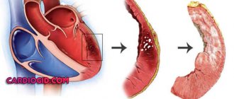
Phlebectasia of the esophagus occurs as a result of problematic blood flow in the hepatic veins. The pathology is provoked by cirrhosis of the liver or blood clots in the organs.
Phlebectasia of the esophagus
Venous stagnation is the main cause of the development of phlebectasis of the esophagus. Among the etiological factors, there are 2 main groups: hepatic and extrahepatic. Phlebectasia is provoked by cirrhosis, hepatitis, fatty degeneration of liver cells due to metabolic disorders, complications of infectious diseases (tuberculosis, syphilis), parasitic lesions, and hereditary pathologies. In addition, bulging of the esophageal veins occurs against the background of disruption of the cardiovascular system, increased blood clotting, compression of blood vessels, inflammation of the lymph nodes near the liver, and connective tissue pathology.
The peculiarity of phlebectasis of the esophagus is the difficulty of making a timely diagnosis. In the early stages, the disease has an asymptomatic course. As it progresses, the elasticity of the venous walls is impaired, they become brittle, resulting in the formation of ruptures, which leads to bleeding.
Phlebectasia of the esophagus goes through several stages:
- first stage: symptoms are not expressed, however, during instrumental examination it is possible to detect an expansion of the venous lumen by several millimeters, foci of ectasia are either absent or appear sporadically;
- the second stage is characterized by the appearance of the first symptoms of phlebectasia; instrumental diagnostics reveal tortuosity of the veins, the walls are enlarged in the lower sections, the mucous membranes are not changed, the vascular lumen is expanded to 1 cm;
- third stage: the veins are thickened by more than 1 cm, fill the cavity of the esophagus by two-thirds, the nodes are detected visually, reflux develops;
- the fourth stage is the most severe, bleeding develops, the mucous membranes are damaged, the nodes are located in clusters, there is no lumen of the esophagus.
The first and second degrees are the initial stages of the development of pathology, which are detected only during an instrumental examination, the third and fourth stages are characterized by distinct signs, requiring immediate initiation of treatment.
Symptoms of esophageal phlebectasis:
- discomfort and chest pain;
- heartburn, belching;
- difficulty swallowing dry food;
- shortness of breath not associated with physical activity.
As the disease progresses, without medical assistance, a dangerous complication may develop - bleeding. Its presence is indicated by the red tint of the vomit (in some cases they take on the color of coffee grounds), feces may become liquid and black, shortness of breath intensifies, the patient feels fatigue, fatigue, heart function is disrupted, and a decrease in the concentration of iron in the blood is detected.
The presence of such symptoms allows one to suspect a pathology of the esophagus, but additional diagnostic tests are necessary to confirm the disease.
Diagnosis of phlebectasis of the esophagus:
- laboratory tests can detect signs of bleeding: a decrease in hemoglobin, the number of red blood cells, occult blood when performing a coprogram;
- with the help of instrumental methods it is possible to identify the cause of the development of phlebectasia: ultrasound of the abdominal organs, esophagoscopy, MRI and CT, radiography of the esophagus with contrast;
After confirmation of the diagnosis, all treatment measures are aimed at preventing bleeding or stopping it. In addition, it is important to eliminate the cause of phlebectasia. The main directions of treatment: surgical and medicinal. In addition, the doctor recommends changing your diet.
If the disease is diagnosed at stages 1-3, conservative therapy is carried out; at stage 4 of the pathology, surgery is indicated.
Drug therapy:
- beta blockers to lower blood pressure;
- vitamin therapy;
- colloidal solutions;
- hemostatic agents;
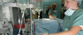
The operation is performed in severe cases when complications have developed and drug treatment is ineffective. One of the surgical techniques is selected:
- electrocoagulation – removal of damaged areas of a vessel using current;
- installation of a bandage - special rubber discs over the damaged vein;
- endoscopic doping - ligation of blood vessels using special attachments or nylon threads;
- sclerotherapy - injection of a special solution into the vessel that glues damaged elements;
- bypass surgery - installation of a stent connecting blood vessels;
- balloon tamponade - compression of blood vessels using a probe.
Classification
Classified according to the location of the disease:
- phlebectasia of the jugular vein;
- esophagus;
- lower extremities.
By origin they are divided into:
- congenital phlebectasia;
- acquired.
Doctors distinguish stages of development based on the clinical picture.
- The first degree is detected during endoscopy by chance - the expansion does not exceed 3 millimeters, the lumens are not darkened, ectasia is not present or is small.
- The second degree is characterized by venous adjustment - strong expansion, for example, if the problem concerns the esophagus, then their volume occupies about a third of the organ cavity.
- The third stage is determined by enlargement and swelling of the veins, the presence of nodule formations, occupies more than half the volume of the esophagus, and there is a deterioration in the condition of the mucosa.
- The fourth stage threatens with thinning of the walls of the mucous membrane, and in other organs there are cluster-shaped formations and bleeding.
The prognosis is favorable in the first 3 stages. The neglected fourth can be fatal, especially phlebectasia of the IJV.
Phlebectasia of the jugular veins
With phlebectasis of the jugular vein, one of the branches is affected: internal, external or anterior. As a rule, the pathology is not dangerous, but is a cosmetic defect. However, with significant damage to the internal jugular veins (IVVs), which drain blood from the skull, complications may develop.
Causes of phlebectasis of the jugular veins:
- injury to the neck, skull, spine with the development of inflammation;
- violation of sterility when installing a catheter or performing injections;
- inflammation in tissues located near the jugular vein;
- penetration of drugs (for example, calcium chloride) into the tissues surrounding the vessels;
- venous congestion in injuries of the back and chest;
- being forced to be in an uncomfortable position;
- cardiovascular pathology associated with decreased elasticity of the vascular wall;
- the presence of neoplasms compressing the vessel.
If symptoms of the disease appear, you should seek medical help. The doctor will refer the patient for additional examination to confirm the diagnosis.
Diagnosis of phlebectasis of the jugular veins:
- Ultrasound;
- MRI and CT;
- phlebography;
- lab tests.
Based on the results of additional diagnostics, treatment is prescribed. Non-advanced stages of the disease are amenable to conservative therapy. Anti-inflammatory drugs in the form of ointments (Ibuprofen, Indomethacin), antihistamines (Suprastin), and broad-spectrum antibiotics in case of bacterial infection are used.
In most cases, surgery is not required, but the patient must visit a doctor periodically to monitor the condition. In case of severe pathology and the risk of complications, one type of operation is performed:
- circumferential resection of extension;
- longitudinal resection;
- strengthening the vascular wall with a polymer mesh;
- resection with vein grafting.
Possible complications
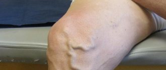
The disease can either manifest itself or occur in a closed, latent form. Tumor formations appear, which increase in size over time and become tense. The skin underneath becomes thinner, and ulcerative processes begin.
This leads to varicose veins, and when squeezing or lifting the limbs upward, the unpleasant symptoms decrease. Phleboliths - round formations - are present in the later stages. The disease progresses slowly, pain, motor dysfunction and trophic problems are possible.
With intestinal pathology, pain occurs in the stomach and liver. As the disease progresses, the symptoms intensify, and the walls of the esophagus and stomach become thinner. This is fraught with disruption of the gastrointestinal tract.
Persistent dilatation of the veins of the esophagus and jugular vein phlebectasia
This circulatory disorder leads to dilation of the superficial veins.
There are various forms of pathology: dilation of the veins of the lower extremities (varicose veins), rectum (hemorrhoids), esophagus, jugular vein.
Phlebectasia of the jugular vein
The jugular vein is several pairs of veins located in the neck and carrying blood away from the brain and neck. There is a pair of internal jugular vein, external vein (includes the posterior auricular vein, occipital and suprascapular) and anterior vein.
Causes
Dilatation of the jugular vein can be observed in both adults and children.
The causes of this disease may be blood stagnation due to injuries, prolonged sitting, or the presence of heart failure.
Provoking factors can also be cancer (leukemia, kidney cancer) and chemotherapy treatment of oncology.
Symptoms of the disease
Often, dilatation of the jugular veins can be asymptomatic.
Some experience a hoarse voice, neck pain, and difficulty breathing. A striking distinctive symptom is a noticeable swelling in the neck. Depending on the type of jugular vein, swelling may vary.
When the external vein expands, small round formations are observed; the internal vein is characterized by a spindle-shaped formation.
Diagnostic techniques
To accurately diagnose phlebectasia of the internal jugular vein (IJV), several studies are performed.
Namely: ultrasound examination, phlebography, puncture, duplex scanning and others, which allow us to present a clear picture of the circulatory system in the neck, back of the head and shoulders.
Healing procedures
Since this disease is very rare, first of all, in the absence of an acute form, the patient is simply observed.
They recommend avoiding stress and prescribing specific medications to eliminate unpleasant accompanying symptoms. After diagnosis and determination of the feasibility of surgical intervention, the dilated vein is surgically removed.
Possible complications
In extremely rare cases, the expansion of the jugular vein can be so strong and large-scale that it threatens rupture, heavy bleeding and subsequent death.
Dilation of the veins of the esophagus
When the blood pressure in the portal vein for a long time exceeds the norm several times, phlebectasia of the esophagus begins. In this case, the outflow of blood is disrupted, the veins accumulate it, change their size and shape.
What causes the disease
Like any venous expansion, dilation of the vessels of the esophagus occurs due to stagnation of blood circulation and increased blood viscosity.
Very often, phlebectasia of the esophagus occurs in parallel or after some other disease, for example: cirrhosis, echinococcosis, amyloidosis, sclerosis, stenosis or thrombosis.
There are cases of congenital varicose veins of the esophagus, this occurs due to dysfunction of the heart.
Signs of pathology
In the initial stages of the disease, the patient may experience regular, light bleeding.
They lead to iron deficiency anemia, hypotension, weight loss, weakened immunity, physical inactivity and weakness. A clear sign of internal bleeding, in this case, is dark stool.
In more serious cases, the patient begins to vomit blood. Some complain of regular belching, heartburn, pain in the epigastric region and behind the sternum.
From the visual side, the patient’s abdomen enlarges due to the large amount of fluid (ascites), dilated veins located on the surface become visible.
Diagnostics
In addition to the patient’s standard complaints, to clarify the diagnosis, the doctor may prescribe a fibroesophagoscopy, which will check the degree of dilatation of the veins, the condition of the blood vessels, the presence of erosive and ulcerative lesions and identify the real cause of bleeding.
X-ray contrast studies may also be used.
Why do legs swell during pregnancy and what methods of prevention exist. In this case, our article will answer whether there is a danger for mother and child.
Anyone prescribed the drug Normoven will benefit from this article #8212; pros and cons, instructions for use and other useful information.
What treatment methods are there?
First of all, they begin to reduce the pressure in the portal system using medication. Peptic esophagitis is prevented or treated with the help of astringents and antacids.
The next step is to remove the dilated vein using bypass or obliteration. As a last resort, surgical removal is performed.
An important part of treatment is the elimination of the cause of phlebectasia, to reduce the likelihood of subsequent dilation of one of the veins of the esophagus.
It is recommended to avoid physical activity, and a strict diet is prescribed, excluding hot, spicy, sour and large-caliber foods.
How dangerous is the disease?
Most often, phlebectasia manifests itself as varicose veins. All other forms of the disease are quite rare. Which in turn has a certain negative imprint on diagnosis and treatment.
In the initial stages of the disease, when it develops slowly, the patient may not even notice abnormalities.
Mild pain, belching, dark stools may either go unnoticed or be attributed to other factors. You can remain in this state for quite a long time, until bloating on the abdomen becomes noticeable and some symptoms worsen.
Therefore, in order to avoid unpleasant surprises, those who have provoking factors (heart failure, circulatory system disorders, sclerosis, thrombosis, etc.) should be regularly examined by their doctor .
In any other case, if there are signs of internal bleeding, unreasonable and regular abdominal pain, you must immediately get checked and, if necessary, undergo a series of examinations to detect the causes of these symptoms.
Perhaps this is something more dangerous than phlebectasia, so you shouldn’t put off going to the doctor for too long.
Help with vein diseases.
Copying of materials is permitted only with indication of the original source.
Join us and follow the news on social networks
https://stopvarikoz.net/other/flebektaziya.html
Manifestations
Pathology of the esophagus occurs at stages 1-2 asymptomatically. The patient is not bothered by pain. Sometimes there is a burning sensation in the chest and shoulder blades, and belching with a sour taste. As the disease progresses, pain symptoms are observed in the chest, esophagus, stomach, and liver.
At stage 4, vomiting of blood occurs, and purple blood clots are present in the stool. Swallowing will be impaired. Constant loss of blood leads to anemia, a person feels weak, cannot work or move normally, loses weight, feels short of breath and has a strong heartbeat.
Phlebectasia in a child is usually diagnosed as congenital. Enlarged and tortuous venous ducts are noticeable and can be localized anywhere.
It appears either from birth or in the first months of life. The color of the skin changes, the integument becomes cyanotic. Next, tumor formations appear.
Phlebectasia or dilatation of the jugular vein in the neck
Marvelous! This is what varicose veins are most afraid of.
It should be applied to the skin where there are swollen veins.
Features of the disease
Another important factor is the discharge of blood from the veins located deep under the muscles into the superficial veins. This non-physiological redistribution of blood, due to a number of reasons, causes dysfunction in the entire venous network, also leading to vasodilation.
Causes
It should be noted that phlebectasia does not depend on the age of the patient; it can equally occur in both an adult and a child.
Causes of dilatation of the jugular vein:
Often, with the development of dilatation of the jugular vein, there are several factors that cause the disease.
Carrying out diagnostics
To identify and make a final diagnosis, a specialist will need the results of several laboratory and instrumental studies:
These are the main diagnostic methods that are used to make a final diagnosis. At the same time, the doctor can prescribe only some of them to obtain a complete information picture of the disease.
However, to identify the exact causes of the disease, it may be necessary to consult specialists who will help determine the main factor in the occurrence of jugular vein phlebectasis. Such specialists include a neurologist, endocrinologist, and oncologist.
Symptoms of the disease
Like any other varicose veins, phlebectasia of the jugular vein initially occurs without any obvious symptoms. If the exposure factor is insignificant, then the disease can develop for years without leaving any traces on the body.
In the future, a feeling of pressure may develop at the site of the expansion of the jugular vein, especially when bending, screaming or sudden movements of the head.
In advanced cases, painful sensations appear in the neck, the voice becomes hoarse, and difficulty breathing may occur.
The last two cases require immediate treatment, since the development of such symptoms negatively affects the general condition of the body.
Treatment methods
After making a diagnosis and recognizing that the jugular vein is dilated, it is time to decide on treatment procedures.
If the dynamics are rapid or the expansion of the jugular vein already has a negative effect on the body, a decision is made to surgically treat the disease. It all comes down to removing the affected area of the vein and connecting healthy areas into one vessel. Also read about vascular surgery for varicose veins
Complications and their prevention
Complications from such conditions are rare. Basically, this is the threat of rupture of the affected and weakened section of the vein and subsequent heavy bleeding. This condition is fatal in most cases.
To prevent this scenario, jugular vein enlargement should be treated whenever possible. If the doctor suggests or even insists on early surgical intervention, it should be performed.
Preventive measures
The main preventive measures can be called:
The main emphasis should be on people who are predisposed to dilation of the jugular vein due to hereditary characteristics.
It must be remembered that vein diseases are difficult to prevent, but you can easily stop and get rid of them in the initial stages of development. That's why regular checkups with your doctor will help you avoid problems in the future.
Are you one of those millions of women who struggles with varicose veins?
Have all your attempts to cure varicose veins been unsuccessful?
Therefore, we recommend reading the story of our reader Ksenia Strizhenko about how she cured varicose veins Read the article
The materials presented are general information and cannot replace medical advice.
https://atlasven.ru/venyi/flebektaziya.html
Diagnostics
If symptoms of pathology are found, contact a physician. Based on anamnesis collection, palpation, and examination, he will make a primary diagnosis and determine the localization of varicose veins.
Laboratory tests include general and biochemical blood tests. It is carried out as prescribed by the therapist.
But it is not always possible to establish an accurate clinical picture and select the correct course of treatment based on laboratory tests and anamnesis.
They also resort to instrumental diagnostic methods:
- endoscopy;
- x-ray;
- scanning of the vascular network;
- esophagoscopy.
Naturally, the principle of collecting tests, prescribing, and complex of examinations change depending on the localization of tumor processes. For consultation, if problems are found in the gastrointestinal tract, contact a gastroenterologist, lower extremities - a phlebologist. In any case, pathology requires a comprehensive examination, which is carried out by several doctors.
First aid for attacks is to stop bleeding. The doctor selects the optimal treatment plan to not only stop it, but also to prevent the next one, which is very likely to happen.
Then drugs that relieve inflammatory processes are included. The purpose depends on the area in which the dilated veins are localized.
Choose:
- antacids, which reduce acidity in the stomach;
- diuretics;
- vasoconstrictors;
- hemostatic drugs.
Beta blockers and vitamin kits are used as therapy. To restore blood flow and blood components, solutions are taken. In some cases, especially if the disease progresses and is at stages 3-4, a blood or plasma transfusion will be required.
In case of severe damage to the venous network, surgical intervention is resorted to. It is carried out using various methods, in particular, electrocoagulation, bandaging, and ligation. In case of cirrhosis, bypass surgery is chosen.
After surgical intervention on the organs of the gastrointestinal tract, the patient is prescribed a lifelong diet that prevents an increase in the acidity of gastric juice.
Features of the disease
Depending on the location, dilatation of the veins of the lower extremities, rectum (hemorrhoids), esophagus and jugular veins are distinguished. Vascular damage occurs under the influence of various factors, including: inflammatory changes, dysfunction of valves, changes in blood flow.
When exposed to one or more pathogenetic factors, an inflammatory reaction is triggered in the vascular wall, phlebitis, periphlebitis, and thrombophlebitis develop. In the presence of microorganisms in the lesion, an infectious process occurs; in their absence, an aseptic process occurs (this form is more often provoked by the influence of drugs).
The expansion of the jugular vein does not depend on the gender and age of the patient; in addition, it can be congenital. Men over 50 years of age are more susceptible to damage to the vessels of the esophagus.
Who's at risk
Adult men (over 50 years old) are at risk. According to statistics, women suffer from phlebectasia 3-4 times less often than men.
Phlebectasia at stage 1 is not dangerous, but at stage 3-4 it leads to consequences. The symptoms are not pronounced and can be confused with other diseases. If there is any suspicion, contact a therapist.
Preventive measures include limiting physical activity, choosing the right diet and taking vasoconstrictor medications.
The material was prepared specifically for the website venaprof.ru, edited by pharmacist M.N. Aleksandrova.