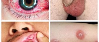
Haemorrhoids
- pathological enlargement of hemorrhoids (internal nodes - internal hemorrhoids, external nodes - external hemorrhoids). Combined hemorrhoids are an enlargement of both external and internal hemorrhoids [1, 4].
The cause of pathological enlargement of hemorrhoids is acute or chronic circulatory disorders in cavernous formations. Along with circulatory disorders, dystrophic changes in the ligamentous apparatus of hemorrhoids play a significant role in the development of hemorrhoids (LE 5, SR D [4, 5]).
Under the influence of these factors, hemorrhoids increase in size, shift in the distal direction, while the processes of degeneration in the retaining apparatus increase, and the hemorrhoids begin to fall out of the anal canal. The development of dystrophic processes in the common longitudinal muscle of the submucosal layer of the rectum and the ligament of Parks, which hold the cavernous bodies in the anal canal, leads to a gradual but irreversible displacement of hemorrhoids in the distal direction.
Publications in the media
Superficial thrombophlebitis is an inflammatory disease characterized by the development of thrombosis and reactive spasm of the superficial veins, most often of the lower extremities. Often develops as a complication of varicose veins. Iatrogenic thrombophlebitis of the superficial veins sometimes occurs after venipuncture or venosection.
Localization . The great saphenous vein and its tributaries are most often affected in the upper third of the leg, lower and middle thirds of the thigh. Rare forms: thrombophlebitis of the superficial veins of the mammary gland and anterior chest wall (Mondor's disease), as well as thrombophlebitis of the dorsal vein of the penis.
Classification . Acute, subacute, recurrent; migratory thrombophlebitis (repeated thrombosis of various parts of the superficial veins, most often in the lower extremities); ascending and descending.
Etiology . Phlebitis can develop independently and cause venous thrombosis, or the infection quickly joins the primary thrombosis of the superficial veins. In addition, it can be secondary to vasculitis, for example, periarteritis nodosa and thromboangiitis obliterans (Buerger's disease). Migratory thrombophlebitis can be a marker of carcinoma, most often of the tail of the pancreas. Thrombophlebitis of the dorsal penile vein is usually caused by acute or chronic trauma.
Pathogenesis . Thrombophlebitis develops in the presence of the following conditions: slow blood flow, increased blood clotting, damage to the wall or valves of the veins, and infection.
Clinical picture • Acute thrombophlebitis occurs suddenly, accompanied by an increase in body temperature followed by chills. Along the course of the affected vein, a painful dense infiltrate in the form of a cord is palpated. Over the infiltrate, hyperemia of the skin with compaction of the subcutaneous tissue is possible. Regional lymph nodes are enlarged • The subacute form of thrombophlebitis occurs without acute local inflammatory phenomena, with normal or slightly elevated body temperature in the first days. When walking, moderate pain, general malaise • The recurrent form is characterized by the emergence of a new area of damage to the superficial veins or exacerbation of a previously occurring process • With Mondor's disease, thrombophlebitis is usually localized in the upper anterolateral part of the mammary gland, or spreads from the lower part of the gland through the submammary fold towards costal arch and epigastrium.
Laboratory tests • Leukocytosis with a shift of the leukocyte formula to the left and increased ESR • PTI • Duplex ultrasound scanning can confirm the spread of a blood clot to the deep veins of the limb • With migratory thrombophlebitis, a thorough examination of the digestive organs is necessary to exclude a primary oncological disease. In diagnostically unclear cases, it is recommended to determine the content of CEAg in the blood serum.
Differential diagnosis • Erysipelas • Lymphangitis • Cellulitis • Tendonitis • Rupture of the medial head of m. gastrocnemius.
TREATMENT
Conservative treatment • Bed rest, elevated position of the limb • Elastic bandage helps fix the blood clot in the superficial veins • Locally: compress with Vishnevsky ointment, semi-alcohol compress, heat • Physiotherapy: iontophoresis with thrombolytin (trypsin-heparin complex) • Drug treatment •• Anti-inflammatory therapy: acetylsalicylic acid, NSAIDs (reopyrin, phenylbutazone). Anticoagulants are usually not prescribed, except in cases of spread of the process to the deep venous system •• Recurrent superficial thrombophlebitis - antibiotics •• Acetylsalicylic acid as a weak anticoagulant •• Troxerutin 0.3–0.6 g/day for 2–4 weeks.
Surgical treatment . The Troyanov–Trendelenburg operation is performed in the presence of a blood clot at the mouth of the great saphenous vein (the blood clot is removed).
Complications • Hyperpigmentation of the skin over the affected vein. • Organized thrombus fixed inside a varicose vein. • Thrombophlebitis of deep veins. About 12% of patients with thrombophlebitis of the great saphenous vein above the level of the knee have spread of the thrombus to the deep veins of the limb (through perforating veins or saphenofemoral anastomosis). • PE • Suppurative phlebitis may be complicated by metastatic abscesses or septicemia • Streptococcal lymphangitis
The prognosis is favorable with timely and adequate appropriate treatment. The patient should be informed that the illness may last 3–4 weeks or longer. With varicose veins of the superficial veins of the lower limb, the likelihood of relapse is very high. To prevent it, phlebectomy is recommended.
Synonym . Thrombophlebitis of superficial veins
ICD-10 • I80 Phlebitis and thrombophlebitis
APPLICATIONS
Thrombosis of the superior sagittal sinus is a blockage of the superior sagittal sinus, the only sinus that carries out venous outflow from the dura mater, with a thrombus. Causes : infection, complication of osteomyelitis, dehydration, trauma, tumors. Clinical picture : fever, headache, lethargy, stretching of the veins of the scalp, convulsions, aphasia, hemiplegia; usually occurs acutely. Treatment depends on the cause: antimicrobials, drainage, surgery. Synonym : thrombosis of the superior longitudinal sinus. ICD-10 . G08 Intracranial and intravertebral phlebitis and thrombophlebitis.
Cavernous sinus thrombosis is a disease characterized by infection of the venous canals that carry out venous drainage from the face and orbit. Causes : direct penetration of infection from the eye socket and face. Risk factors : diabetes and immunodeficiency states. Clinical picture : exophthalmos, swelling of the optic nerve nipple, headache, convulsions, septic fever. The prognosis is unfavorable. Treatment : Hospitalization, intensive care, intravenous fluids and antibiotics. Synonym : cavernous sinus syndrome. ICD-10 . G08 Intracranial and intravertebral phlebitis and thrombophlebitis.
Renal vein thrombosis . Causes : abdominal surgery, chronic heart failure, dehydration, kidney disease, decreased renal blood flow. Clinical picture : lumbar pain, dysuria, enlarged painful kidney. The course can be acute or chronic. Treatment : anticoagulants, glucocorticoids. ICD-10 . N28 . 0 Renal ischemia or infarction.
Thrombophlebitis - symptoms and treatment
Therapeutic and preventive measures for thrombophlebitis are complex and can be conservative and surgical. The main objectives are to maximize the elimination of risk factors, reduce and alleviate local symptoms in acute thrombophlebitis, prevent the spread of thrombophlebitis to the deep vein network and prevent venous thromboembolic complications.
Surgical treatment
Not long ago, the gold standard for the treatment of ascending thrombophlebitis was crossectomy (Troyanov-Trendelenburg operation), but practice results have shown that this method of surgical intervention is the most traumatic and life-threatening for patients.
When the process goes beyond the sapheno-femoral or sapheno-popliteal anastomosis, thrombectomy is performed from the main veins. Surgery can be performed using regional anesthesia or intubation endotracheal anesthesia. The preference for thrombectomy method depends on the level of location of the proximal part of the thrombus.
In case of perforator thrombosis, thrombectomy from the perforator vein is performed. In case of embolic thrombosis of the femoropopliteal segment, ligation of the superficial femoral vein (SFE) is indicated.
In case of embolic-dangerous iliocaval thrombosis, plication of the inferior vena cava is performed.
The figure shows the implantation of a vena cava filter into the inferior vena cava, the indication for which is embolic iliocaval thrombosis.
Laser treatment
According to the latest revised clinical guidelines, endovenous laser coagulation (EVLC) is a low-traumatic and safe technique for ascending thrombophlebitis. This method can be used to operate on any category of patients. As a rule, surgery is performed under local tumescent anesthesia.
Conservative treatment
Today, for existing indications, the most effective method will be anticoagulant therapy. In medical practice, it is customary to distinguish between direct-acting anticoagulants, which help reduce thrombin activity in the blood, and indirect-acting anticoagulants, which prevent the formation of prothrombin in the liver. Low molecular weight heparins belong to the group of direct-acting anticoagulants. These include medications such as Enoxaparin sodium (Anfibra, Clexane, Hemapaxan, Lovenox), Dalteparin (Fragmina) or Tinzaparin, which must be administered subcutaneously 1-2 times during the day. The use of low molecular weight heparins results in maximum effectiveness and minimal side effects. Indirect anticoagulants are Warfarin derivatives, which require special caution and a high degree of laboratory control (INR). Currently, the greatest interest is in drugs that do not require laboratory monitoring of INR and with lower risks of complications, for example, such as Xarelto (Rivaroxaban) or Pradaxa.
In addition, the patient is prescribed long-term wearing of compression hosiery and auxiliary pharmacotherapy, phlebotonic drugs (Detralex; Venarus; Phlebodia 600), etc. It is also advisable to prescribe non-steroidal anti-inflammatory drugs and local treatment.
Treatment of thrombophlebitis of superficial veins and acute thrombophlebitis
Recently, international consensus has accepted the equality between the terms “acute thrombophlebitis” and “thrombophlebitis of the superficial veins”, which determines the commonality of pathogenetic mechanisms, complications and treatment tactics.
Physiotherapeutic treatment
Intermittent pneumocompression is a physiotherapeutic method of massaging tissue using special multi-chamber cuffs with different operating pressures. This technique has excellent lymphatic drainage properties, helps reduce swelling and can be used for thrombophlebitis of the veins of the lower extremities.
Electromyostimulation using the VENOPLUS device - this patented technique consists in the fact that electromyostimulation leads to muscle contraction and activation of the muscle-venous pump. It can also be used for thrombophlebitis of the veins of the lower extremities.
Gymnastics for thrombophlebitis
This disease poses a threat to life if a blood clot breaks off, so gymnastics is contraindicated.
Leeches for thrombophlebitis
Currently, the first line of therapy is the use of low molecular weight heparins. Hirudotherapy has a much lower effect.
Ointments for thrombophlebitis
The use of ointments is possible in the complex treatment of thrombophlebitis. NSAIDs are used in the form of ointments to relieve pain. Such ointments include Fastum Gel, diclofenac, ortofen and others. It is also possible to use ointments with heparin (Lioton, hepatrombin).
Nutrition for thrombophlebitis
Nutrition does not affect the course of thrombophlebitis. It is worth adjusting the diet only if the disease occurs against the background of obesity.
Can folk remedies help?
Decoctions, tinctures and compresses have no proven effectiveness and therefore cannot be recommended for the treatment of thrombophlebitis. Thrombophlebitis is a disease that can threaten the patient’s life and therefore requires immediate and adequate treatment.
Rehabilitation
Rehabilitation usually takes place at home. Compliance with any special regime is not required. If the patient smokes, he should stop smoking if possible.
Surgery of venous and lymphatic pathology
Vein diseases
The body's venous system consists of an extensive network of veins - vessels through which blood moves from organs and tissues to the heart and lungs. When moving from the lower extremities to the heart, the blood flow overcomes the force of gravity. The function of the pump that pushes blood up through the veins is performed by the muscles that contract when walking, and the valves on the inner lining of the veins prevent the return flow of blood. Defects of the venous valves play a significant role in the development of venous diseases: varicose veins and its consequences - chronic venous insufficiency, trophic ulcers, thrombophlebitis.
Phlebology is a specialized branch of cardiovascular surgery that studies the anatomy, physiology of blood circulation, as well as diseases of the venous vessels, issues of their prevention, diagnosis and treatment. Treatment of vein diseases is carried out in departments of vascular pathology or phlebology centers. Vein diseases today are one of the most common diseases of the vascular system; these are diseases of young people of working age, mainly from 25 to 45 years. Untimely treatment of venous pathology is fraught with dire consequences, including disability and death.
Symptoms of venous disease that require immediate medical attention include fatigue, heaviness, cramps and pain in the legs, swelling, vascular dilation in the legs, the appearance of areas of discoloration and thickening of the skin and subcutaneous tissues.
To assess the degree of venous circulation disorder and select further treatment tactics, diagnostics are carried out using duplex ultrasound scanning and Doppler ultrasound - modern high-precision methods for studying the condition of blood vessels.
In the treatment of venous pathology, conservative methods (drug and compression therapy), non-surgical endovasal methods (laser coagulation, sclerosing therapy of veins) and surgical treatment methods are used.
In modern phlebology, low-traumatic methods for the treatment of vein diseases, based on the use of endoscopic, laser, and radiofrequency technologies, have become widespread, allowing to minimize complications and the rehabilitation period after the intervention. Timely treatment can achieve a good cosmetic result, which is especially important for women suffering from diseases of the veins of the lower extremities.
To prevent the development of diseases of the veins of the lower extremities, phlebologists advise following simple but very effective rules.
- Avoid prolonged standing or sitting in a cross-legged position.
- Avoid compressing the blood vessels of the legs with tight clothing and elastic bands, wearing high heels
- To improve venous outflow from the lower extremities during rest, it is necessary to give the legs an elevated position relative to the body
- To strengthen the leg muscles, you should perform self-massage, engage in physical therapy, and take a contrast shower.
- Avoid hot baths, frequent visits to baths, saunas, and excessive tanning.
- To reduce blood viscosity and the tendency to form blood clots, increase fluid intake to 2 liters per day
- For the initial manifestations of varicose veins, as well as for the purpose of its prevention during pregnancy, it is recommended to use medical compression hosiery
Varicose veins of the lower extremities (International Classification of Diseases, 10th revision (ICD-10) / IX Diseases of the circulatory system (I00-I99) / I80-I89 Diseases of the veins, lymphatic vessels and lymph nodes, not classified elsewhere / I83 Varicose veins lower extremities) is an important medical and social problem due to the tendency towards an increase in morbidity in people of working age, an increase in the number of complicated forms and, as a consequence, the formation of permanent disability. Varicose veins are characterized by high prevalence. So in Russia, according to the most rough estimates, it is diagnosed in 30 million people. In the USA and Western European countries, about 25% of the population suffers from various forms of varicose veins. After 70 years, the disease occurs in more than 70% of people. Mostly women over the age of 20 are affected (5 times more often than men). Clinical picture The main clinical symptoms of varicose veins are as follows:
- visible dilated veins;
- fatigue of the lower extremities;
- a feeling of fullness in the calf muscles, burning, soreness in the lower extremities;
- periodic and later permanent swelling of the feet and legs;
- night cramps in the calf muscles;
- general bluishness of the skin of the lower extremities;
- age spots, etc. (see pictures).
The severity of the symptom complex is greater in the evening, after physical exertion, and in hot weather. Stages of development In the development of varicose veins of the lower extremities, 4 stages are distinguished: 1st stage (compensation). There are minor cosmetic defects (spider veins), no complaints. 2nd stage. Twisted dilated veins appear, slight swelling of the ankles, mild night pain. Stage 3 (subcompensation). Reporting, night cramps in the calves, rapid fatigue of the legs, a feeling of muscle swelling, and skin pigmentation are observed. Stage 4 (decompensation). Severe swelling of the feet and ankles, a sharp increase in the width of the veins, acute pain, itching, severe cramps. Signs of thrombophlebitis and venous ulcers often appear.
Varicose veins of the lower extremities are one of the most common diseases on earth. The only radical, pathogenetically substantiated method of treatment is surgery. Conservative therapy is an addition to the main treatment.
Drawing. Physiology of venous outflow and pathophysiology of varicose veins n/c Acute venous thrombosis (ICD - I80 Phlebitis and thrombophlebitis; I82 Embolism and thrombosis of other veins) is the formation of a “blood clot”, a thrombus, in the lumen of a vein (more often observed in the deep venous system of the lower limbs, much less often - upper).
Drawing. Venous thrombosis.
Risk factors for venous thrombosis are, for example, young women taking oral contraceptives, fractures of limb bones, mass formations of the pelvis and retroperitoneal tissue, prolonged bed rest, paraplegia, the postpartum period, cancer, however, it is difficult to identify specific causes in a significant number of cases. The pathogenesis of venous thrombosis consists in the acute occurrence of an obstacle to venous outflow and redistribution of blood along collaterals against the background of inflammation of the venous wall; after which the process (period) of recanalization of thrombosed veins and restoration of pathological blood flow through them begins, which (period) ends by the sixth month. But even in recanalized veins, the blood flow does not become normal because the lumen of the vein does not reach its original diameter, and the destruction of the valves after thrombosis leads to retrograde blood flow. The consequence of venous thrombosis is Postthrombophlebitis syndrome (PTPS) or postthrombotic disease. Diagnosis of acute venous thrombosis in outpatient and inpatient settings is based on an assessment of clinical symptoms and the results of instrumental examination methods and should be carried out as soon as possible since the clinical prognosis depends on the speed of determining the fact of thrombosis, its localization, and the nature of the proximal part of the thrombus. In all cases of acute venous thrombosis, it is preferable to begin the examination with ultrasound angioscanning.
Drawing. Ultrasound angioscanning (ultrasound of n/c veins).
Treatment Goals
venous thrombosis: stop the spread of thrombosis, prevent thromboembolism of the pulmonary arteries, prevent the progression of edema and thereby prevent possible venous gangrene and limb loss, restore the patency of the veins (prevention of post-thrombophlebitis disease, prevent relapse of thrombosis. Suspicion of deep vein thrombosis of the lower extremities, especially if established diagnosis are an indication for emergency hospitalization of the patient (if possible, in a specialized angiosurgical hospital, in extreme cases in a general surgery department). The main objective of surgical treatment is to prevent pulmonary embolism. For this purpose, when an embolic (floating) thrombus is identified, depending on the specific clinical situation, direct or catheter thrombectomy, percutaneous implantation of vena cava filters of various designs, ligation or plication of the great veins/inferior vena cava.In all other cases, treatment problems are solved with the help of conservative therapy. Also, conservative therapy must be carried out after any of the listed surgical interventions.
Drawing. Scheme: a) completed operation of thrombectomy from the femoral veins supplemented by plication of the SMV according to the original method of the Bakulev Center; b) front view; c) side view; d) in cross section. Anticoagulant therapy is indicated for all patients with clinical and laboratory signs of active thrombus formation, which usually corresponds to the first three weeks of the disease. This is the most effective means of stopping the progression of thrombosis with a proven therapeutic effect. Anticoagulant therapy should be carried out with mandatory consideration of contraindications to these drugs. Anti-inflammatory drugs (NSAIDs) are used due to the fact that there is inflammation of the venous wall and perivasal tissues, as well as pain syndrome, which makes it difficult for the patient to activate. Local treatment includes local hypothermia in the projection of vascular bundles, as well as the use of ointments (gels are preferable), the main active principles of which are heparin and NSAIDs. You should not use warming alcohol and ointment compresses, which can support the phenomena of phlebitis and contribute to the progression of thrombosis.
Non-drug treatment methods: adherence to a certain motor regimen and elastic compression. The more carefully the patient follows the motor regimen and compression therapy regimen in the acute stage of the disease and during the rehabilitation period, and the longer the time it is carried out, the better the results of treatment of venous thrombosis, and the less pronounced the phenomena of chronic venous insufficiency in the long-term postthrombotic period.
Postthrombophlebitic syndrome (PTFS) or postthrombotic disease (ICD - I87 Other venous lesions / I87.0 Postthrombotic syndrome) is a chronic obstruction of venous outflow from the lower extremities, developing after deep vein thrombosis. Clinically, PTFS can manifest itself several years after acute thrombosis. Patients experience a bursting feeling in the affected limb and painful night cramps, ring-shaped pigmentation and swelling are formed, which over time acquires fibrous density. Diagnosis of PTFS is based on anamnestic data and ultrasound results of the veins of the lower extremities. Increasing decompensation of the venous circulation is an indication for surgical treatment.
With thrombosis, a blood clot forms in the lumen of the vein. After the acute process subsides, the thrombotic masses are partially lysed and partially replaced by connective tissue. If lysis predominates, recanalization occurs (restoration of the vein lumen). When replaced by connective tissue elements, occlusion occurs (disappearance of the lumen of the vessel). Restoration of the vein lumen is always accompanied by destruction of the valve apparatus at the location of the thrombus. Therefore, regardless of the predominance of one or another process, the outcome of phlebothrombosis is a persistent disturbance of blood flow in the deep vein system. Increased pressure in the deep veins leads to dilation (ectasia) and incompetence of the perforating veins. Blood from the deep vein system is discharged into the superficial vessels. The saphenous veins dilate and also become incompetent. As a result, all veins of the lower extremities are involved in the process. Blood deposition in the lower extremities causes microcirculatory disorders. Impaired skin nutrition leads to the formation of trophic ulcers.
Classification There are two variants of the course (edematous and edematous-varicose forms) and three stages of postthrombophlebitic disease.
- transient swelling, “heavy leg syndrome”;
- persistent swelling, trophic disorders (skin pigmentation disorders, eczema, lipodermatosclerosis);
- trophic ulcers.
Symptoms The first signs of PTFS may appear several months or even years after acute thrombosis. In the early stages, patients complain of pain, a feeling of fullness, heaviness in the affected leg when walking or standing. When lying down and placing the limb in an elevated position, the symptoms quickly decrease. Modern research in the field of clinical phlebology shows that in 25% of cases, PTPS is accompanied by varicose veins of the superficial veins of the affected limb. Edema of varying severity is observed in all patients. A few months after the development of persistent edema, inductive changes in soft tissues appear. One of the characteristic signs of PTFS is ring-shaped pigmentation, which begins above the ankles and covers the lower third of the lower leg. Subsequently, dermatitis, dry or weeping eczema often develop in this area, and in the later stages of the disease, poorly healing trophic ulcers appear. The course of PTFS can be different. In some patients, the disease manifests itself with mild or moderately severe symptoms for a long time, while in others it rapidly progresses, leading to the development of trophic disorders and permanent disability. Diagnostics. If post-thrombophlebitis disease is suspected, the doctor finds out whether the patient suffered from thrombophlebitis. When collecting anamnesis, it is necessary to pay attention to episodes of severe prolonged swelling and a feeling of fullness of the affected limb. To confirm the diagnosis, an ultrasound scan of the veins of the lower extremities is performed. To determine the shape, location of the lesion and the degree of hemodynamic disturbances, direct or computer radiopaque venography is used.
Treatment of PTFS. Conservative therapy During the adaptation period (the first year after thrombophlebitis), patients are prescribed conservative therapy. The indication for surgical intervention is early progressive decompensation of blood circulation in the affected limb. At the end of the adaptation period, treatment tactics depend on the form and stage of PTFS. In the stage of compensation and subcompensation of circulatory disorders, constant wearing of elastic compression devices and physiotherapy are recommended. Even in the absence of signs of circulatory disorders, patients are contraindicated in hard work, work in hot shops and in the cold, and work associated with prolonged standing on their feet.
In case of circulatory decompensation, the patient is prescribed antiplatelet agents, fibrinolytics, and drugs that reduce inflammation of the vein wall. For trophic disorders, pyridoxine, multivitamins, and desensitizing agents are indicated.
Surgery. Surgery cannot completely cure a patient with PTFS . The operation only helps to delay the development of pathological changes in the venous system. Therefore, surgical treatment is performed only if conservative therapy is ineffective.
The following types of operations for PTFS are distinguished:
- reconstructive interventions (vein resection and plastic surgery, bypass surgery);
- corrective operations (phlebectomy and miniphlebectomy - removal of dilated saphenous veins, ligation of communicating veins).
Forecast. To date, no type of treatment, including surgical interventions, can stop the further development of the disease if its course is unfavorable. Within 10 years from the moment of diagnosis of postthrombophlebitic disease, disability occurs in 38% of patients. I89 Other non-infectious diseases of the lymphatic vessels and lymph nodes/ I89.0 Lymphoedema, not elsewhere classified
Lymphatic pathology Lymphedema (elephantiasis) (ICD-10: I89.0; I97.2; Q82.0) is a polyetiological disease associated with impaired lymphatic drainage, which causes disruption of protein metabolism in tissues, progressive and irreversible formation of fibrous tissue, leading to significant enlargement and deformation of a limb or any part of the body. The reasons that lead to these changes may be associated with a congenital anomaly of the lymphatic vessels or be a consequence of their damage of various types.
Drawing. Patient M., 23 years old, with stage III congenital lymphedema of the left leg. A) before treatment and B) 4 months after resection, skin-plastic surgery.
Diseases of the venous system of the extremity, in particular postthrombotic disease, are not the cause of the development of lymphedema, but if the patency of the deep veins is impaired, secondary persistent disorders of lymph circulation in the extremity of the “pseudoelephantiasis” type are often observed.
Lymphedema is a common socially significant disease. According to WHO, about 10% of the population suffers from this pathology. More than 10 million people worldwide suffer from lymphedema caused by chronic infection. Patients with lymphedema account for 2.5-7% of all patients with peripheral vascular disease. The main group of patients are young and middle-aged women. The number of newly identified patients increases every year, and there is no downward trend in the incidence rate. The social significance of the topic under discussion is great and is due to the fact that the majority of patients are people of working age (96%), for whom rehabilitation is extremely important.
How does lymphedema develop? Lymphedema can be congenital or acquired. In the first case, it can appear in early childhood or during puberty, when hormonal levels change. But congenital pathologies are less common. Lymphedema often occurs as a complication after skin diseases, oncology and gynecological problems.
Drawing. Patient B., 62 years old, with stage III postmastectomy lymphedema of the left upper limb. Lymphedema tends to constantly progress. In areas of edema, skin changes, trophic disorders (ulcers, dermatitis, eczema), recurrent erysipelas, and fungal skin lesions (mycosis) begin to occur. The last stage of the disease is called “elephantiasis” due to the strong increase in size of the limb. It is important to stop the development of the disease, to eliminate existing problems at the initial stages of the process, when irreversible changes have not occurred in the skin and subcutaneous tissue.
Lymphedema is a chronic, progressive disease; it cannot be cured forever , but lymphedema can be significantly reduced or completely eliminated with the help of comprehensive treatment.
Clinical manifestations of this disease are: swelling of the upper or lower extremities, impaired nutrition of the skin, the appearance of trophic changes in the skin and subcutaneous fat. The most common complications of lymphedema are erysipelas, hyperkeratosis, papillomatosis, and lymphorrhea.
Diagnosis of lymphedema is based on the patient’s complaints, anamnesis of the disease and life, and features of the clinical picture. To confirm the diagnosis, special instrumental research methods are used:
- lymphoscintigraphy (radioisotope study of lymphatic vessels);
- magnetic resonance and computed tomography (allows visualization of tissues and organs layer by layer if an oncological etiology of lymphedema is suspected);
- lymphography.
- imfovenous anastomoses
- Vascularized lymph node transplantation
The choice of a rational method of treatment for lymphedema of the extremities is determined by the clinical course, stage of the disease and extent of the lesion. Treatment should be comprehensive, individual, with constant preventive measures. Currently, conservative and surgical methods are used, each of which has its own indications. The main goal in the treatment of lymphedema is to restore the outflow of lymph from tissues and reduce lymphedema. Conservative therapy as the main method of treatment is indicated for patients with stage I lymphedema. For more severe lymph circulation disorders, the method is auxiliary and is carried out when preparing patients for surgery and as a preventive course after surgical treatment.
Drawing. Patient S., 47 years old, with stage IIb-III lymphedema of the lower extremities before (photo on the left) and after (photo on the right) a course of inpatient complex anti-edematous therapy (20 bed days according to compulsory medical insurance standards). Reduction in the circumference of the left shin by 25 cm and hip circumference by 7 cm.
Among the surgical methods used:
Drawing. Scheme and intraoperative photo of lymphovenous anastomosis. The operation is performed under a microscope using microsurgical techniques and instruments.
Lymphorrhea (lympha - moisture, rhoe - flow), or lymphorrhagia, is a fairly rare pathological condition. May be traumatic or non-traumatic in nature. The traumatic form of lymphorrhea is more often diagnosed in young and middle-aged people. The non-traumatic variant of the pathology is detected mainly in patients of the middle and older age groups. Minor losses of lymph do not pose a danger to the body. With massive single lymphorrhagia or chronic leakage, exhaustion develops and death is possible.
Causes of lymphorrhea. Lymphorrhagia is a polyetiological condition that occurs during injuries, operations, and some diseases. The main causes of pathology are considered:
- open and closed injuries to lymphatic vessels of significant diameter, most often the thoracic duct;
- accidental injury to vessels or manipulation of such vessels during operations in certain anatomical areas;
- rupture of altered vessels with lymphangiectasias, vascular tumors, blockage by cancer emboli, especially against the background of lymphostasis and lymphangitis.
Classification Along with the outflow of chylous fluid, the following variants of lymphorrhea are distinguished: lymphocele - lymph accumulates in the tissues; chylothorax - fluid is detected in the pleural cavity; chylopericardium – lymph flows into the pericardial cavity; chyloperitoneum - fluid is found in the abdominal cavity. The release of lymph with urine due to its leakage into the urinary tract is called chyluria; leakage through the intestinal wall leads to the development of exudative enteropathy.
Diagnostics Depending on the cause of the development of lymphorrhea, thoracic surgeons, oncologists, cardiologists and other specialists can diagnose this pathology. The diagnosis of external lymphorrhagia is made on the basis of anamnesis, complaints and the results of a physical examination. In case of internal loss of lymph, additional research is necessary. Diagnosing external lymphorrhea is usually not difficult. Lymphorrhagia in the body cavity must be differentiated from the accumulation of other fluids. Chylothorax is distinguished from hydrothorax, pleurisy, hemothorax. Chyloperitoneum is differentiated from various variants of ascites, chylopericardium is differentiated from hemopericardium and exudative pericarditis.
Treatment of lymphorrhea The patient is hospitalized in a hospital according to the profile of the underlying disease. Treatment tactics are determined by the localization of lymphorrhagia.
NMITs SSH im. A.N. Bakuleva of the Ministry of Health of the Russian Federation has unique experience in performing all kinds of operations on the main veins and lymphatic system. We operate on patients with varicose veins of the lower extremities, perform reconstructive interventions for PTF, venous aneurysms and compression venous stenoses. To date, at the National Medical Research Center of Sports Sciences named after. A.N. Bakulev Ministry of Health of the Russian Federation performs about 100 operations on the lymphatic system per year (the largest number of operations among clinics in the Russian Federation). The Center performs almost all types of surgical interventions existing in the world for congenital and acquired pathologies of the great veins and lymphatic system, the results of which correspond to the world indicators of the world's leading clinics specializing in such operations.