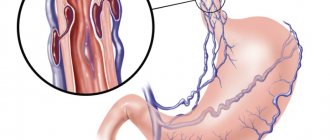
An optical apparatus and the diagnostic method of fibrogastroduodenoscopy help determine the amount of blood loss recorded in the organs of the upper gastrointestinal tract. This is facilitated by the classification of bleeding according to Forest. This gradation, as you will see, is quite simple and convenient. In particular, it helps doctors notice all pathological changes in blood vessels (both venous and arterial), determine the stage of the disorder, and the volume of blood that the patient loses. We will talk to you in more detail about gastric bleeding and their classification later.
What causes such bleeding?
Before we look at Forest’s classification of bleeding, let’s talk in more detail about them themselves.
What is the cause of bleeding in the upper gastrointestinal tract? Several points can be highlighted here:
- Internal organ injuries.
- Intoxication of the body.
- Inflammatory processes.
- Damage to the gastrointestinal tract by infection.
- Ulcer or tumor of the stomach, esophagus or duodenum.
- A rarer cause is hereditary pathologies affecting blood vessels and blood. In particular, Rendu-Osler disease, hemophilia.
Additional Research
In addition to FGDS, the gastroenterologist will need other data about the patient’s well-being. A clear diagnosis will be established after several step-by-step studies. The doctor needs the readings to complement each other and create an overall picture.
A comprehensive study of the whole organism is necessary.
- Study of the patient's blood composition. A biochemical blood test is needed.
- Composition of urine.
- Medical history (anamnesis), as well as information about the presence of other diseases.
- MRI diagnostics of all internal organs.
If lethargy, pallor, and clouding of consciousness are observed - these are clear signs of blood loss, the patient’s blood pressure and hemodynamics of the heart must be monitored. Those diagnosed with any heart disease undergo fibrogastroscopy under the supervision of an experienced cardiologist.
Main symptoms
It is important for the patient to know not only the Forest classification of bleeding, but also the symptoms of this pathological condition. These will include the following:
- Blood streaks that will be observed in the vomit.
- Uncharacteristic shade of vomit. Most often the color of coffee grounds.
- Loss of strength, chronic feeling of weakness.
- Constant feeling of thirst.
- Low blood pressure.
- Dizziness.
- “Flies are flying before my eyes.”
- Pale skin.
- Tachycardia.
- Black stool.
Danger to the patient
Along with Forest's classification of bleeding, many are interested in how dangerous this pathological condition is for humans. If such blood loss occurs for the first time in a patient, then statistics will show a small risk - 10% of cases are fatal. But recurrent bleeding in the upper gastrointestinal tract is already dangerous. There are 50% of deaths from all diagnosed cases. Moreover, the age of the patients does not have a significant impact on this figure.
However, it is noted that the risk of bleeding in the upper gastrointestinal tract is most typical for older people. The reason is that they are more likely to develop serious complications of any pathological processes in the body. And most often it is the upper parts of the digestive system that are affected - this is 70% of all cases.
Diagnostics
Forest's classification of gastric bleeding is used by doctors specifically when diagnosing such cases. The doctor begins his work with the following:
- The patient's medical history is collected. The doctor analyzes the symptoms of the pathological process.
- First of all, laboratory methods are prescribed to study the patient’s health status - a general analysis of a blood sample, a study of its coagulability.
- The patient is prescribed FGDS - fibrogastroduodenoscopy. If a person is admitted to the hospital with gastric or intestinal bleeding in critical condition, then this diagnostic method will be the only one for him.
Forest's classification for gastrointestinal bleeding will explain the examination results obtained using the FGDS technique. Its recognized effectiveness is confirmed by several factors:
- Helps document an episode of gastrointestinal bleeding.
- Determines the location of the damaged vessel.
- Sets the intensity of bleeding. And this helps specialists understand how much blood a person has lost.
Another plus is the convenience of the procedure. It is performed both at the patient’s bedside and in a specialized treatment room.
Analyzing the endoscopic classification of bleeding according to Forest, I would like to draw the reader’s attention to another useful function of FGDS. The technique allows you to influence the defect of a “leaky” vascular wall:
- clipping;
- coagulation;
- fibrin filling;
- injecting the damaged area with drugs that stop bleeding.
How is diagnosis carried out?
If a person is suspected of having gastric bleeding, specialists use an integrated approach to diagnose it.
That means:
- collecting anamnesis, that is, history, disease;
- identifying early signs of gastric bleeding;
- laboratory testing of blood to determine its clinical indicators (primarily, determining the level of hemoglobin and studying the properties of coagulation);
- conducting instrumental examination using, first of all, endoscopic methods (FGDS).
Carrying out fibrogastroduodenoscopy (FGDS) allows you to establish the presence of gastric bleeding, determine the location of the damaged vessel and the degree of intensity of blood loss.
Contraindications for fibrogastroduodenoscopy
The Forrest classification of ulcerative bleeding is thus applied only on the basis of diagnostic data obtained by fibrogastroduodenoscopy. This gradation cannot explain the results of other studies.
Contraindications to EGD therefore make it impossible to use the classification for abnormal gastrointestinal bleeding in an individual patient. These include the following:
- Stroke.
- Acute stage of myocardial infarction.
- State of agony.
Indications for use
Indications for performing FGDS are the presence of the following diseases in the patient's medical history:
- peptic ulcer of the stomach and/or duodenum;
- malignant forms of cancer, in which the disintegration of neoplasms is observed;
- erosive gastritis;
- Randu-Osler syndrome (hereditary pathology in which defective blood vessels are formed);
- oncological and genetic pathologies of the circulatory system.
Categorical contraindications for endoscopic examination are the acute period of myocardial infarction, stroke, and a state of agony.
Classification of bleeding according to Forest, its meaning
This classification was developed by Dr. Forest back in 1974. It was intended for the gradation of bleeding, which was established during fiberoscopy of the gastric region and duodenum. The basis for the classification was clinical studies that were directly conducted by Forest himself.
The significance of gradation today is that it has introduced into medical practice that unified system for drawing up a protocol, which is done by a specialist after each FGDS procedure. In particular, this is a technique for visualizing a pathological focus of bleeding. It is important to highlight that it helps to choose the most appropriate treatment tactics for the patient, which directly affects the speed and quality of his recovery.
Representation of Forest classification
Let us present to readers the classification of ulcerative bleeding according to J. A. F. Forrest.
The first group is F1 (bleeding in the active stage). Within it, two additional categories will be distinguished:
- F1a - in this case, blood will flow out of the damaged vessel in a stream (sometimes in a pulsating stream).
- F1b - here the blood mass flows out of the vessel drop by drop. Another name is vessel sweating.
The second group is F2. In these cases, the bleeding has already stopped. The group also has its own subcategories:
- F2a - the device detects a thrombosed vessel at the bottom of the ulcer. Diameter - less than 2 mm.
- F2b - a fixed blood clot was found at the bottom of the ulcer. The diameter here will be more than 2 mm.
- F2c - black spots are visible at the bottom of the ulcer. This is what a network of small, already thrombosed vessels looks like.
The last group is F3. There are no subcategories here - this is how the specialist will indicate the result of the examination, in which no gastric or intestinal bleeding was detected (the bottom of the ulcer is clean).
Thus, the classification makes it possible to determine both the stage of pathological bleeding and its activity. The peculiarity of the gradation is that it helps to choose the right therapy for a particular patient:
- If blood loss is observed, the specialist first takes measures for hemostasis (stopping bleeding).
- If vascular bleeding has stopped, then help to the patient is aimed at replenishing the volume of fluid lost by his body, restoring normal blood counts, blood pressure, and pulse.
- If bleeding was not detected, then this fact indicates either the absence of defects in the vascular wall or the healing stage.
Gastric resection
If type F1a is established - a pulsating stream, blood quickly fills the gastrointestinal tract. In some cases, self-medication should not be allowed. You need to call an ambulance immediately. Giant ulcers, which are more than 2.5-3 cm in diameter, and peptic ulcers are operated on urgently.
The tactics of surgeons when preparing for surgery consists of three points:
- Forrest group definitions.
- Carrying out early endoscopy, which determines the level of damage to the mucosa and the degree of risk.
- Determining the volume and timing of the operation. It is determined what kind of resection is needed: distal resection, proximal, or gastrectomy.
In cases where the ulcer is dangerous and there is a risk of repeated intense bleeding, a planned distal gastrectomy is prescribed. The operation is not dangerous. During this procedure, 2/3 of the organ, the pylorus, is removed. The remaining stump is connected to the small intestine or duodenum. The operation is possible only after the bleeding has completely stopped.
Total resection or gastrectomy with resection is the removal of more than 90% of the organ tissue. Prescribed after endoscopy when repeated ulcerations were observed, large volumes of blood were lost, and pyloric stenosis was found.
Further treatment
The diagnostic results allow you to choose the optimal treatment method for a particular patient:
- Conservative. This is drug therapy.
- Operational. This is a surgical intervention of various specifications and scope - from excision and suturing of the ulcer itself to resection of the gastric region.
Today, as progress develops, Forest's classification is used in various modifications and variations. However, the entire system of grading gastrointestinal bleeding is still aimed at determining the activity of the pathological process and selecting the best treatment option for the patient. Classification helps to identify the likelihood of relapse of the pathology, which can be fatal for the patient.
Advantages of the method
The advantage of the FGDS method is the ability to carry it out almost anywhere - both in a specialized office and in a department ward. Based on the results of the examination, having classified the bleeding according to Forest, the specialist can decide on the tactics of further treatment measures - either prescribe conservative methods of treatment, that is, using hemostatic drugs, or immediately refer the patient to the surgical department.
The use of endoscopic methods also makes it possible to determine the volume of necessary surgical intervention - suturing a damaged vessel, excision of an ulcerated area of the mucosa, resection of the stomach. Under endoscopic control, it is possible to directly influence the damaged vessel wall - coagulation, clipping, fibrin filling, injecting the damaged area with hemostatics.
The procedure for fibrogastroduodenoscopy

