Thoracic osteochondrosis
is a degenerative-dystrophic disease in the middle parts of the spine. It occurs much less frequently than lumbar and even cervical osteochondrosis and accounts for about 10% of all cases of the disease. Symptoms of thoracic osteochondrosis are associated with several features of the spine in the thoracic region: rigid fixation of the vertebrae due to the ribs and skeletal muscles and less mobility, a semicircular bend of the ridge in this region and the corresponding distribution of the load.
Thoracic osteochondrosis is a disease of the middle parts of the spine.
Symptoms of thoracic osteochondrosis begin with a violation of tissue trophism and dehydration of the intervertebral discs. Lack of nutrients and fluid leads to the fact that cartilage loses its elasticity, begins to crack and “sag”
.
This state of affairs is fraught with compression of the nerve roots, deterioration of sensitivity and functioning of internal organs, as well as severe pain. Often, the destruction of the vertebrae in the thoracic region is accompanied by the appearance of osteophytes
- bone outgrowths that injure the paravertebral tissues and lead to chronic inflammation.
Contrary to popular belief, thoracic osteochondrosis is not a natural age-related process and requires special treatment. The onset of the disease can occur at the age of 25 years
;
approximately 70% of the total number of patients are women aged 35 to 55 years. Since the disease begins long before retirement age, it can lead to limitations and even loss of ability to work
.
Unlike other types of osteochondrosis, thoracic osteochondrosis may not manifest itself for a long time - or disguise itself as other pathologies, which is why it is called a chameleon disease. It is also the most difficult to treat. Therefore, it is very important to promptly monitor the symptoms of thoracic osteochondrosis.
- and today we will tell you which ones.
Causes of heart pain
Acute aching pain in the heart area occurs in a person regardless of age, conditions and circumstances. However, not every one of them may indicate heart problems. Depending on the mechanism of formation, all chest pains can be divided into cardiac and non-cardiac etiologies.
Stitching pain in the heart area can occur for various reasons, with diseases of other organs with irradiation of the pain syndrome into the chest.
- Diseases of the neuromuscular system:
- Thoracic osteochondrosis;
- Pain in the cervical spine;
- Myalgia;
- Intercostal neuralgia.
- Pathologies of large vessels:
- Aortic aneurysm;
- Pulmonary embolism.
- For pathology of the digestive system:
- For stomach diseases and heartburn;
- Pancreas;
- Esophagus;
- Gallbladder.
- Diseases of the respiratory system:
- Bronchial asthma;
- Pneumothorax;
- Pneumonia;
- Pleurisy;
- Tuberculosis.
- Nervous system diseases:
- Vegetative-vascular dystonia;
- Panic attacks.
- Diseases of viral etiology:
- Shingles.
If, with pain in the heart area, there is pressure and radiates to the left hand and little finger, this may result in angina pectoris. Acute pain syndrome develops with myocardial infarction, dull pain characterizes chronic ischemic heart disease.
Treatment of thoracic osteochondrosis and its prevention
Conservative treatment of osteochondrosis in the thoracic spine is intended to stop or at least slow down degenerative changes, restore normal back mobility and eliminate symptoms that cause discomfort to the patient.
Therapeutic treatment of thoracic osteochondrosis involves the simultaneous use of:
- medications
(chondroprotectors, neuroprotectors, muscle relaxants, anti-inflammatory drugs, analgesics); - methods of physiotherapeutic complex
; - therapeutic exercises
; - orthopedic regime
.
Patients are also advised to change their diet and lifestyle.
In case of severe irreversible changes in the intervertebral joints , in which pain and nerve conduction disorders are not relieved by medications,
surgery is recommended for patients
. It helps stop the death of nerve tissue and prevent life-threatening or disabling consequences of thoracic osteochondrosis. Depending on the situation, complete or partial resection of the intervertebral disc or its replacement with an artificial one, narrowing of the spinal canal or other surgery may be indicated.
Treatment of thoracic osteochondrosis
Physiotherapy
The objectives of physiotherapy for osteochondrosis of the thoracic region are to reduce pain and inflammation, relieve spasms, strengthen the muscle corset, restore the function of the nerve roots and normal blood circulation.
To relieve the symptoms of thoracic osteochondrosis, the following are successfully used:
- Magnetic therapy
is one of the most effective anti-inflammatory techniques. Improves metabolic processes in tissues and relieves swelling. - Laser therapy
. Promotes biological activation of regenerative processes. Helps eliminate the consequences of trophic disorders and relieve inflammation. - Drug electrophoresis
. Allows you to restore tissue nutrition and relieve inflammation - the effect of the procedure depends on the medications used. - Medicinal phonophoresis
. Ensures deep penetration of the active ingredients of medications into soft tissues. - Massotherapy
. Helps relax muscles, eliminate tension and improve the supply of nutrients to the tissues of the spine. In the early stages, it eliminates the main symptom of thoracic osteochondrosis - a feeling of pain in the sternum - in a few sessions. - Acupuncture
. Stimulation of muscles and nerve endings helps relieve pain, restore sensitivity and eliminate swelling. - Ultrahigh frequency therapy
. Increases the permeability of capillary walls, improves blood flow and ensures the flow of protective cells into the site of inflammation. - Shock wave therapy
. It starts the processes of restoration of bone and cartilage tissue, prevents the deposition of calcium salts on the vertebral surfaces. - Balneotherapy
. As a rule, mud and ozokerite applications are used, less often paraffin. Radon and hydromassage baths are also recommended for patients. They help improve metabolism and restore sensitivity in affected tissues. - Amplipulse therapy
. It has a neurostimulating, analgesic and trophic effect, activates metabolic processes, and facilitates breathing. - Kinesiotherapy
(physical therapy, massage, traction therapy, kinesiotaping). Allows you to strengthen ligaments and skeletal muscles, restore mobility in the back and eliminate even persistent spasticity. Prevents the formation of osteophytes and narrowing of the canals in which the spinal roots are located.
In addition to physical therapy sessions, for the treatment of thoracic osteochondrosis, patients may be recommended an orthopedic corset, which allows them to relieve the load on the spine.
Exercise therapy and massage
Therapeutic exercises and massage help strengthen the back muscles and relieve stress on the spine. With daily sessions, they help achieve stable drug-free remission, increase range of motion, and eliminate neurological manifestations of the disease. These treatment methods also prevent complications of osteochondrosis.
. For example, congestion in the lungs (with thoracic osteochondrosis it is difficult to breathe deeply), due to which patients are susceptible to pneumonia, as well as coronary heart disease.
Dosed physical activity helps relieve compression of the nerve roots, improve blood circulation and nutrition of the intervertebral discs. The optimal frequency and duration of gymnastic classes is determined by the exercise therapy instructor. As a rule, 3-4 exercises of 10-15 minutes a day are enough.
.
The exercises recommended for the treatment of thoracic osteochondrosis include the following:
- Stand straight, feet together, hands at your sides. As you exhale, raise your arms up and bend back, then inhale deeply. Lower your arms and lean forward, slightly arching your back in a dome-shaped manner (to do this, lower your head and shoulders as you exhale).
- Sit on a chair and, while inhaling, place your hands behind your head. Bend back and rest your shoulder blades on the back of the chair, exhaling.
- Get on all fours and arch your back. After maintaining the position for 3 seconds, bend your back with a crampon.
- Lying on the floor on your stomach, place your palms on the floor and, raising yourself on your arms, try to move your head as far back as possible, lifting your chest off the floor.
- Lie on your stomach and extend your arms at your sides. Perform the “yoke” exercise, trying to simultaneously raise your head and legs.
- Sit on the floor and stretch your legs in front of you. Reach the fingers of your right hand to the toe of your left foot and vice versa.
- Do a plank exercise (about 30 seconds).
- Perform hangs on the horizontal bar (or, in the absence of a horizontal bar, secure your fingers on the door frame and try to stretch your back as much as possible).
Bends to the side while raising your arm will also be helpful. All exercises should be performed 8 to 10 times
.
To treat thoracic osteochondrosis, various massage techniques are used, incl. acupressure and vacuum massage. For self-massage at home, patients are recommended the following movements:
- stroking the cervical-collar, scapular and axillary areas
; - squeezing with the thumb and index finger
(grabbing the skin and soft tissues); - trituration
; - kneading
(it should be done extremely carefully, it is advisable to entrust this technique to a specialist).
Massage should not be performed during exacerbations of the disease or during severe inflammation.
Drug treatment of thoracic osteochondrosis
To treat the symptoms of thoracic osteochondrosis, the following groups of drugs are used:
- Nonsteroidal anti-inflammatory drugs (NSAIDs)
- help relieve pain, calm inflammation and disperse swelling. They can be used both systemically (in the form of tablets, capsules and injections) and locally (in the form of ointments, gels, creams, compresses and solutions for medicinal electrophoresis). - Hormonal drugs
- to eliminate acute and chronic neurological pain. Used in cases where NSAIDs demonstrate insufficient effectiveness. - Muscle relaxants
are prescribed to reduce muscle tone and eliminate spasms of skeletal muscles. This helps alleviate pain and has a positive effect on tissue trophism. - Blood circulation correctors
- strengthen the walls of blood vessels and improve blood circulation in small capillaries that nourish the periosteum and other structural elements of the vertebral joints. Reduce discomfort and reduce the risk of complications. - Neuroprotectors
- to preserve and restore sensitivity during compression of nerve roots and relieve neurological symptoms. This group also includes cholinesterase inhibitors, which improve nerve impulse transmission and help restore normal muscle tone.
For structural restoration of tissues affected by the degenerative process, the following are used in the treatment of thoracic osteochondrosis:
- chondroprotectors (Artracam)
are essential bioactive substances that trigger the processes of regeneration of cartilage and bone tissue. Serves to prevent the growth of osteophytes and narrowing of the spinal canal. They help a weakened body grow stable and resilient cartilage cells. Taking Artracam significantly improves the shock-absorbing properties of intervertebral discs, making them more elastic and resistant to damage; - vitamin complexes
- help normalize metabolic processes and prevent excess oxidation in tissues.
To relieve excruciating pain that interferes with the patient’s daily activities, the doctor may recommend a medical blockade with anesthetics. Diuretics are used to eliminate swelling and relieve compressed nerves and blood vessels.
Prevention of thoracic osteochondrosis
To prevent symptoms of thoracic osteochondrosis, you should:
- Take care to maintain healthy posture. This is facilitated by walking, swimming and therapeutic exercises to strengthen the muscles of the back, chest, abs, and shoulder complex.
- When working sedentarily, properly arrange your workplace and perform physical warm-up whenever your back and neck begin to become stiff (ideally, bend to the sides, stretch, and rotate your shoulders every 2 hours).
- Avoid back injuries and promptly seek help from an orthopedic traumatologist, even in the case of a seemingly insignificant bruise. Other diseases of the musculoskeletal system should not be triggered, especially in the joints of the lower extremities.
- If possible, unload the spine during the day (to do this, just lie on the floor for 40 minutes).
- Protect yourself from hypothermia and stress.
- Take chondroprotectors to protect joints for at least 3 months a year. Regardless of the strength of the muscular corset, the human spine is anatomically not adapted to vertical loads, and therefore requires additional support.
- Adjust weight if it is overweight. In addition to maintaining a low-carb diet, you should eat a diet high in vitamins and minerals. In the spring-autumn period they can be taken in tablet form.
- Sports activities that involve jumping from heights or lifting heavy weights should be limited.
- It is advisable to sleep on a semi-rigid bed, and for prolonged sitting, choose hard furniture. This helps maintain muscle tone and relieve stress on the spine. If possible, you should purchase an orthopedic mattress and shoe insoles.
- Do not lift loads weighing more than 10 kg. The load should be distributed evenly, with muscle tension and without transferring to one side. If necessary, use a special sports corset. It is undesirable to hold loads in outstretched arms for a long time.
- Women should avoid wearing high-heeled shoes. The optimal heel height is 2-4 cm.
These recommendations will also be useful for those who are already sick - they will have a beneficial effect on the condition of the spine and will help to significantly slow down pathological changes.
And remember: the main thing in the prevention of thoracic osteochondrosis is consistent adherence to healthy habits.
Let chest pain never bother you!
What are the symptoms of pain in different diseases?
Often, pain in the heart area when inhaling is attributed to purely heart diseases, without even suspecting that the real reason lies in something completely different.
How the pain syndrome manifests itself depends on the causes and level of progression of the disease. For example, with heartburn, a burning sensation also occurs in the sternum area. They occur when gastric juice enters the esophagus. Heartburn is also characterized by the appearance of belching and an unpleasant sour taste. These symptoms appear after eating, when bending the body, or in a lying position. In this case, antacid medications are prescribed for relief. When heartburn occurs and pain radiates to the heart area, the cause does not always lie in the esophagus.
- These may be symptoms of gastroesophageal reflux disease (GERD), which is also characterized by heartburn;
- Spasm of the esophagus with disruption of muscle function, which leads to problems with swallowing and moving food in the stomach;
- With achalasia, this pathology manifests itself in disruption of the valve between the stomach and esophagus, with food retention and the appearance of pain in the chest.
Depending on the nature of the pain in the heart area, inflammatory diseases can be diagnosed:
- Gall bladder with the development of cholecystitis;
- Pancreas and acute manifestations of pancreatitis;
- Gallstone disease (GSD).
Painful symptoms in these diseases can radiate into the chest cavity and resemble heart pain.
Pulmonary diseases can also be accompanied by sharp and dull pain in the chest. These signs can occur with pneumonia, pleurisy , with characteristic inflammation of the lungs and tissues that line the chest cavity, respectively. These diseases are characterized by increased pain when inhaling, the appearance of a cough, and an increase in temperature to critical values. Also, diseases can accompany each other and after pneumonia complications arise, which manifest themselves in the form of pleurisy.
Diseases of the respiratory system
Pathologies of the pleura and pleural cavity, as well as the lungs themselves, can provoke pain in the chest. A distinctive feature of these diseases is the presence of a connection between pain and breathing: it increases with inhalation and decreases with exhalation. This causes an involuntary decrease in breathing rate - it becomes frequent and superficial. Most often, chest pain in such situations is caused by pneumonia, pleurisy, pleural empyema and tumors of the bronchopulmonary localization and pleura.
Pneumonia
Pneumonia is inflammation of the lungs that occurs as a result of damage to the lung tissue by a variety of bacteria, viruses or fungi. Most often, it is a consequence of acute respiratory infections, including infection with a new strain of coronavirus, which is accompanied by a deterioration in the patient’s condition after a temporary improvement.
As a result, pneumonia has typical symptoms characteristic of most respiratory infections:
- rapid increase in body temperature to 38 °C and above;
- chills;
- headache;
- decreased appetite;
- cough is initially dry, and then with sputum, less often hemoptysis;
- dyspnea;
- superficial or deep pain in the chest, which tends to intensify with coughing and breathing;
- muscle weakness, joint discomfort;
- general weakness, depression.
Today, diagnosing pneumonia is not difficult and is usually detected during the initial examination of the patient.
Pleurisy
Pleurisy is inflammation of the pleural layers covering the surface of the lungs and chest wall. This is accompanied by the formation of fibrinous deposits or the accumulation of liquid exudate in the cavity formed by two layers of the pleura. This may be a consequence of an infectious lung infection, in particular tuberculosis, although it also occurs with bacterial, viral and fungal infections of other origins. Also, pleurisy can develop against the background of rheumatism, after myocardial infarction, as well as with gastrointestinal pathologies and in some other cases.
There are dry and exudative pleurisy, and the second form is often a consequence of untimely treatment of dry pleurisy. In such cases the following are observed:
- intermittent chest pain, aggravated by deep breathing, coughing, swallowing, and in some cases, by tilting the body in the opposite direction;
- pain radiating to the stomach, shoulder;
- severe, prolonged hiccups;
- lag of the affected half of the chest when breathing;
- dry cough;
- dyspnea;
- weakness, decreased performance;
- increase in body temperature to subfebrile values;
With pleurisy, there is often a friction noise between the layers of the pleura, which resembles the creaking of snow underfoot. Moreover, if you place your palm on your chest, you can feel their friction.
With exudative pleurisy, pain is present at the beginning of the development of the disease and at the end. But after a sufficient volume of exudate accumulates in the pleural cavity, they disappear. The remaining symptoms are generally similar to those of dry pleurisy.
Empyema of the pleura
Pleural empyema, in fact, is one of the variants of the course of pleurisy, but unlike other forms, it is characterized by the accumulation of pus in the pleural cavity. It can be a consequence of pneumonia (pneumonia), the formation of tumors, cysts, bronchiectasis and other disorders of the lungs, less often in the abdominal organs.
The pathology is characterized by:
- chest pain;
- dyspnea;
- fever;
- chills;
- decreased appetite;
- forced position of the body (lying on the sore side).
The development of pleural empyema requires hospitalization in the surgical department.
Tumors of the lungs and pleura
There are many types of benign and malignant neoplasms that can affect the lungs, bronchi and pleura. They are often accompanied by recurrent pleurisy with constantly increasing pain on the affected side. But direct chest pain caused by a neoplasm, especially a malignant one, occurs after it reaches a large size. This significantly worsens the prognosis, although today there are methods for treating even large tumors that were previously considered inoperable.
How to help with acute pain in the heart?
It is no coincidence that when pain appears in the heart area, they do not know what to do until the doctor arrives. Call the hotline number +7 (863) 226-18-17 , which is best entered into the phone’s memory in advance. Experienced private medical doctors help in emergency situations and transport the patient to the hospital. Emergency medical care is provided in clinics, hospitals and at home.
Before the doctor arrives, you should stop moving and take a lying position, and be sure to call a specialized ambulance. If your heart hurts, self-medication can end sadly and lead to death.
In case of high or low blood pressure, take the tablet as indicated. The patient is prescribed sedative drops , Corvalol, motherwort, valerian, and nitroglycerin. If the pain has passed, there is no need for nitroglycerin; it is also contraindicated for low blood pressure. You can put validol under your tongue. Treatment of cardiac pathologies is prescribed by a doctor according to indications.
How to relieve pain under the right shoulder blade
If pain in the scapula area is associated with diseases of the spine, muscles or injury, then painkillers should be taken as first aid. For this purpose, non-steroidal anti-inflammatory drugs or NSAIDs are most often used. They effectively fight pain of any origin, suppressing inflammatory reactions. However, uncontrolled and long-term use of NSAIDs increases the risk of side effects of this group of drugs, such as stomach ulcers, gastric bleeding or kidney failure. Therefore, without a doctor’s prescription, it is permissible to take analgesics for no more than 5 days, after which you must consult a doctor for diagnosis and treatment.
Local remedies in the form of distracting and analgesic ointments, gels and patches do not have such side effects, since this dosage form is limited to the area of application. They help especially well with myositis, muscle strain, attacks of neuralgia and other diseases with chronic pain syndrome. In diseases of the spine, pain can be relieved by rest, limiting physical activity and prohibiting heavy lifting.
It is strictly forbidden to use painkillers if you suspect acute cholecystitis, pancreatitis and other diseases of the abdominal organs that require emergency surgery. Taking painkillers in such patients can lead to temporary relief of symptoms and a false sense of improvement, which ultimately contributes to late presentation and serious condition of the patient due to extensive inflammation of the peritoneum or peritonitis.
If pain in the area of the shoulder blade on the right occurs against the background of severe abdominal pain, accompanied by fever, nausea, and vomiting, you must seek medical help as quickly as possible.
Shpidonov Gennady Stanislavovich
Neurologist
Rostov State Medical University (neurology)
10 years of experience
What diseases cause pain in the heart?
Acute pain syndrome can also appear with pulmonary diseases such as bronchial asthma and pneumothorax. Asthma attacks cause pain throughout the chest cavity. An equally severe form and painful condition is characterized by pneumothorax , in which air penetrates from the outside into the chest cavity and the lungs can collapse with the appearance of pain.
Among the main causes of painful attacks in the sternum area is also pathology of the peripulmonary vessels. They intensify when inhaling with the formation of a cough with pulmonary embolism , pulmonary hypertension with increased pressure in the vascular system supplying blood to the lungs.
Painful vascular diseases include dissecting aortic aneurysm with characteristic damage to large vessels. This emergency condition can be life-threatening. In this case, the pain is initially felt in the heart area and gradually moves down the abdomen. The patient's blood pressure drops sharply, tachycardia occurs and he loses consciousness.
A classic example of the appearance of pain in the chest area is osteochondrosis , which occurs in the thoracic and cervical spine. The pain is reminiscent of angina attacks with radiation to the left shoulder blade and arm. The difference lies in the intensification when moving, raising arms, turning the head, bending the body.
Sharp stabbing pain is characteristic of intercostal neuralgia, Tietze syndrome . It occurs at the joints and spaces between the ribs and becomes more intense when inhaling. The patient cannot take a deep breath, shallow breathing is observed. In case of pathology, strong anti-inflammatory and painkillers are prescribed.
An extremely painful condition in the heart area is characterized by inflammation of the muscle fibers of the chest and back . It resembles similar acute pain in intercostal neuralgia and osteochondrosis.
Shingles , which occurs as a result of infection of the body with the herpes virus, is no less painful In this case, the nerve endings are affected, characteristic rashes appear on the skin, which becomes sensitive, and this whole clinical picture is accompanied by acute pain in the chest.
Painful attacks can occur during panic attacks, nervous disorders of various etiologies. This category of patients is often young, with an unstable nervous system, and in a depressed state after stress. In this case, pain can appear spontaneously or be systematic.
Treatment methods for intercostal neuralgia
Treatment of intercostal neuralgia is complex. The doctor solves several problems simultaneously.
It is necessary to reduce the severity of the pain, and preferably completely eliminate the pain syndrome. For this purpose, anti-inflammatory therapy is prescribed.
It is important to eliminate the cause of the nerve damage. If the disease is viral in nature, antiviral therapy is carried out. For muscular-tonic syndrome, muscle relaxants are prescribed. If a nerve is pinched at the point where it exits the spinal canal, manual therapy may be prescribed. For tumor processes, surgical treatment is necessary.
Treatment is also carried out to strengthen the nerve tissue.
Treatment during periods of acute pain
During periods of acute pain, bed rest should be observed for at least several days. The bed should be flat and firm. “Dry heat” helps a lot. For example, you can wrap a woolen scarf around your chest. Warm compresses, pepper plaster, and mustard plasters are used (it is necessary to avoid placing mustard plasters directly on the spinal column). Massage with anti-inflammatory and warming ointments is recommended. Sedatives and non-steroidal analgesics are used as prescribed by the doctor.
During this period, it is necessary to avoid physical activity and stress. You can't drink alcohol.
Subsequent treatment
Further treatment includes:
- physiotherapy (methods such as UV therapy, UHF, electrophoresis, darsonvalization, magnetic therapy are used);
- massage;
- reflexology;
- physical therapy.
Make an appointment Do not self-medicate. Contact our specialists who will correctly diagnose and prescribe treatment.
Rate how useful the material was
thank you for rating
How to understand that your heart hurts?
Pressing sharp pain in the heart occurs with angina pectoris or “angina pectoris,” which is the main precursor to the development of coronary heart disease (CHD). It radiates to the shoulder, left hand and little finger, and can radiate to the jaw. The intensity increases with nervous tension and physical activity. It occurs as a result of coronary vascular spasm and insufficient oxygen supply to the heart muscle due to atherosclerosis. The pain may go away on its own with rest and rest.
Among the most dangerous heart diseases associated with the cardiovascular system of the body is myocardial infarction . It occurs as a result of blockage (thrombosis) of a coronary artery by an atherosclerotic plaque and cessation of blood supply to an area of the heart muscle. In this case, necrosis occurs and part of the muscle dies.
Pain during myocardial infarction causes severe weakness, lack of air, radiates to the left arm, encircles the shoulder and spreads under the left shoulder blade, sometimes to the neck, jaw, and can form far from the heart area. Sometimes the temperature rises. The person breaks into a cold sweat, shortness of breath, and in some cases nausea.
Chest pain occurs in the case of cardiac pathologies of inflammatory etiology, these include endocarditis , myocarditis , pericarditis. They appear after illnesses caused by viral and bacterial infections.
How is differential diagnosis carried out?
If chest pain occurs, you should immediately call an emergency doctor. Before the specialist arrives, if possible, relieve emotional and physical stress, calm down, lie down and understand the nature of the pain in order to properly notify the doctors.
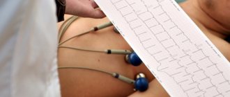
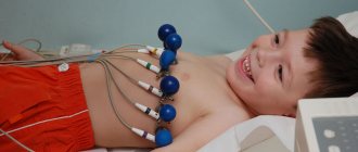
The doctor's examination includes consultations with specialized specialists, instrumental studies, and an electrocardiogram. It shows how the heart works, detecting signs of a heart attack depending on the stage and location.
Medical care and consultations can be prescribed by various specialists, a neurologist, cardiac surgeon, infectious disease specialist, pulmonologist, surgeon, gastroenterologist, psychiatrist. In case of nervous experiences, you may need the help of a psychologist.
Instrumental studies
In addition to an electrocardiogram (ECG), in case of cardiac pathology, various instrumental diagnostic studies .
- Ultrasound of the abdominal cavity;
- FEGDS examination of the digestive organs;
- Ultrasound of the heart, pulmonary vessels and aorta;
- FVD, examination of external respiration functions;
- Chest X-ray;
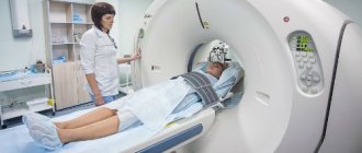
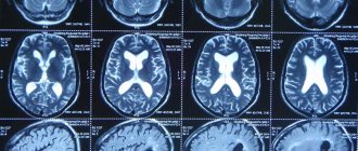
- CT, computed tomography;
- MRI, magnetic resonance imaging;
- EchoCG, echocardiography and others.
Treatment of pain on the right side of the shoulder blade
Pain treatment must begin with diagnosis, since effective pain management is only possible if you know its cause. Symptomatic therapy based on the use of analgesics does not treat the cause of pain, but only temporarily alleviates the most severe symptom. After making an accurate diagnosis, it is possible to treat the underlying disease by influencing the factors that led to it.
This is also important because without knowing the cause of the pain, it is difficult to choose the right treatment. Thus, attempts to treat pain in the scapula on the right side with analgesics in acute cholecystitis can lead to complications in the form of gangrene of the gallbladder, its perforation and extensive peritonitis, with a high risk of death. Painkillers are strictly contraindicated for acute surgical diseases of the abdominal organs.
What signs confirm heart pain?
Critical conditions are caused by cardiac pathologies, which can be determined using simple manipulations. It is necessary to understand whether the pain intensifies when inhaling, when raising your arms, bending your torso, or whether you can take a deep breath. An increase in pain may indicate the presence of intercostal neuralgia or osteochondrosis.
The pain may intensify with physical exertion and decrease at rest when angina pectoris , with insufficient blood supply to the heart muscle in an active state.
Cardiac pathologies are often accompanied by sharp pressing pain, difficulty breathing, stress, discomfort in the chest, cold sweat, irradiation to the left shoulder, arm and under the left shoulder blade.
An emergency call to a private medical ambulance allows you to help in emergency situations, do an ECG, a rapid blood test for the protein troponin to determine myocardial infarction, and transport the patient to the hospital.
Rate this article
Article rating 4.25 out of 5. Votes: 69.
Latest articles
Sexual transmission of infections
Sexually transmitted infections or STIs are dangerous diseases that can undermine health and lead to dire consequences. Most of the diseases in this group are…
Hormonal disorders
A woman’s well-being largely depends on the endocrine system. Any hormonal imbalance can cause a sharp deterioration in health, weakening the immune system...
Cervical dysplasia
Cervical intraepithelial neoplasia or cervical dysplasia is a precancerous condition expressed by pathological changes in cells. In most...