DOI: 10.18508/endo1911
Authors: K.V. Raskina1, Yu.E. Poteshkin2
Information about the authors: 1First Moscow State Medical University named after. THEM. Sechenov, Clinic of Endocrinology, 119435, Russia, Moscow, Pogodinskaya st. 1-1. , 2LLC "Actual Medicine", 129515, Russia, Moscow, st. Academician Koroleva, 13-1-IV/1.
Authors: KV Raskina1, YE Poteshkin2
Authors affiliation: 1 I.M. Sechenov First MSMU, Department of endocrinology, 119435, Pogodinskaya str. 1-1, Moscow, Russia. , 2LLC “Relevant medicine”, 129515, Akademika Koroleva Str. 13-1-IV/1, Moscow, Russia.
Abstract:
In addition to all well-known functions such as the long bones growth stimulation in childhood and lipolysis activation, growth hormone contributes greatly to the development of a healthy heart. Growth hormone (GH) affects the growth of cardiac muscle and its ability to contract. GH activates the production of insulin-like growth factor 1 (IGF-1) cells by cardiomyocytes and vascular endothelium. GH and IGF-1 affect both the structure of the heart and vascular smooth muscle cells. Both together and separately, these hormones can increase the vascular tone and, consequently, strengthen the resistance of the peripheral bloodstream. The use of GH and IGF-1 in the heart failure treatment is being studied. But we cannot omit the fact that an inadequate dosing regimen of GH and IGF-1 can lead to hypertrophy of the left ventricular wall, which leads to the risk of heart ischemia.
- Growth hormone (GH) affects the growth of the heart muscle and its ability to contract.
- GH activates the production of insulin-like growth factor 1 (IGF-1) by cardiomyocytes and vascular endothelial cells.
- Both together and separately, GH and IGF-1 are able to increase vascular tone and, consequently, increase peripheral resistance of the bloodstream.
- With age, the concentration of GH and IGF-1 decreases.
Mechanisms of the influence of GH and IGF-1 on blood vessels
- Regulation of peripheral vascular resistance Intravenous administration of IGF-1 reduced blood pressure within minutes in an experiment on rats
- In a study of healthy people, a single injection of IGF-1 increased cardiac output but had no effect on blood pressure.
- In people with chronic heart failure (CHF), IGF-1 increased (↑) cardiac output and decreased (↓) peripheral vascular resistance
- A recent study achieved a similar result in healthy people: increased blood flow in the forearm along with dilation of resistive vessels.
- To determine the effect of a long-term decrease in IGF-1 in the blood, studies were conducted on transgenic mice: a decrease in circulating IGF-1 led to a significant increase in blood pressure.
- Several options for the mechanisms of the vasodilatory effect of IGF-1 have been proposed: activation of the nitric oxide (NO) system in the endothelium
- release of eicosanoids by endothelial cells
- activation of Na+-K+-ATPase in vascular smooth muscle cells
- increase in the number of ATP-dependent K+ channels in vascular smooth muscle cells
- In people with growth hormone deficiency, compared to healthy people, peripheral sympathetic impulses are increased, affecting vascular smooth muscle cells. One study showed that after 1 year of growth hormone replacement therapy, this impulse is significantly reduced, which leads to a decrease in peripheral vascular resistance.
- A number of large studies have reported hypertension in adults with GH deficiency without replacement therapy.
- Long-term elevation of GH levels results in cardiomegaly, increased cardiac output, and decreased peripheral vascular resistance.
Growth hormone, IGF-1 and the heart
The presence of receptors for GH and IGF-1 in the heart (especially on the membranes of cardiomyocytes) has been discovered by several research groups [3, 4]. Experiments on animals have shown that with an increase in the size of the heart or pressure in the ventricles, the content of IGF-1 in the myocardium increases [5-7]. This is especially noticeable in the left ventricle (LV), probably due to greater stress on its walls [5]. With an increase in pressure in the LV (with aortic stenosis) and its volume (aortic regurgitation), the synthesis of IGF-1 in the human myocardium increases [8]. With increasing load on the ventricular walls, the concentration of mRNA in cardiomyocytes increases biphasically. The direct effects of IGF-1 (but not GH) on the isolated heart and/or on cardiomyocytes have been demonstrated by a number of authors and include increased protein synthesis, increased contractility and changes in calcium flux [9, 10]. In isolated cardiomyocytes, IGF-1 induces early and prolonged expression of the troponin I and myosin light chain-2 genes; At the same time, cell hypertrophy is observed [11]. Finally, administration of GH and IGF-1 to rats with experimental heart failure is accompanied by activation of myocardial growth, dilation of peripheral vessels and changes in cardiac function [12–14].
An increase in heart size and dilation of peripheral blood vessels are the most consistent results of GH administration to humans, while changes in cardiac function are not always recorded [15, 16].
Extensive evidence suggests that activation of the GH–IGF-1 system alters myocardial contractility independently of the growth-promoting effect. U. Vetter et al. [17], and later N. Freestone et al. [10] showed that IGF-1 dose-dependently enhances the contraction of isolated cardiomyocytes. These data were confirmed in isolated perfused heart and papillary muscle [9]. The increase in contractility, according to various studies, is approximately 20-25% of the original. The reaction occurs within just a few seconds after adding IGF-1 to the perfusion medium.
In heart failure, diabetes mellitus, arterial hypertension, and also in the aging process, a kind of “IGF-1 resistance” is observed, which is probably associated with changes in nitric oxide (NO) production [18, 19]. However, using cardiomyocytes from dogs with heart failure, S. Kinugawa et al. demonstrated preservation of the ability of IGF-1 to increase contractility [20].
GH has no direct effect on the contractility of the isolated heart, papillary muscles or cardiomyocytes [9, 11]. It is likely that GH receptors in the heart mediate other effects of the hormone.
The acute inotropic effect of IGF-1 is recorded in all studies, regardless of the model system and animal species. However, there is controversy regarding the subcellular mechanisms of this effect.
Effect of GH and IGF-1 on peripheral vessels
One of the most consistent biological effects of GH is peripheral vasodilation. This effect is believed to be mediated by IGF-1, which has high-affinity binding sites on endothelial cells. IGF-1 directly stimulates NO release, as shown in cultured human endothelial cells [21]. In healthy volunteers, administration of IGF-1 caused a noticeable dilation of forearm vessels, which was reversed by the addition of L-NG-monomethylarginine [22]. NO is a known mediator of vasodilation, an inhibitor of platelet aggregation, leukocyte adhesion and smooth muscle cell growth [23].
The subcellular mechanisms of IGF-1 action on smooth muscle cells include stimulation of the sodium-potassium pump in the reverse mode, which compensates for the release of sodium from the intracellular environment. This results in a decrease in calcium concentration with concomitant muscle relaxation and vasodilation. IGF-1 also increases the release of NO in smooth muscle cells with a subsequent acceleration of cAMP synthesis and a decrease in intracellular calcium concentrations [23].
A study by J. Timsit et al. for the first time suggested that GH improves muscle contractility and at the same time saves energy. In all likelihood, this is explained by an increase in the number of corresponding enzymes and the conversion of the myosin phenotype [24]. An increase in function without an increase in energy costs was also observed when IGF-1 was added to an isolated heart: with an increase in contractility by 20-25%, there were no changes in the concentration of macroenergetic phosphates in the cells [9].
Effect of recombinant GH (rGH) on the heart in humans in the absence of somatotropic insufficiency
Subcutaneous administration of rGH at a dose of 9.6 IU/day for 1 week reversibly increased heart rate (HR), LV systolic function, and cardiac output without concomitant LV wall thickening. Similar, but more pronounced, changes in cardiac function were noted in patients with short-term active acromegaly and in experimental animals [25].
Thus, even a short-term increase in GH levels leads to a state of peripheral hyperperfusion and cardiac hyperkinesis. Over time, this condition can lead to CVS pathology.
Effect of GH deficiency on the heart
Epidemiological data indicate a reduced life expectancy in adult patients with hypopituitarism and an increase in their mortality from cardiovascular diseases by more than 2 times [26, 27]. The cause of increased mortality in patients with hypopituitarism is considered to be GH deficiency. However, cardiovascular risk in GH deficiency is associated not only with the direct effects of GH and IGF-1 on the heart and endothelium, but also with associated changes - abdominal obesity, low physical activity, insulin resistance, increased levels of total cholesterol and low-density lipoprotein cholesterol (LDL). ), atherosclerosis, hypercoagulation, pulmonary dysfunction [28, 29]. Abnormal lipid profiles have been reported to correlate with the severity of GH deficiency [30], as well as an inverse correlation between IGF-1 and LDL cholesterol levels [31].
A number of studies have reported higher blood pressure (BP) levels in patients with GH deficiency than in healthy individuals. The incidence of arterial hypertension among 1000 patients was higher than the population level (22 and 15%, respectively) [32]. In another study, increased sympathetic nervous system activity in patients with somatotropic insufficiency was associated with higher diastolic blood pressure (10 mmHg higher than controls) [33]. A. Colao et al. in a group of 56 patients with GH deficiency, no such dependence was revealed, and at the peak of physical exercise, patients had a lower level of systolic blood pressure [34].
Structural features and functions of the heart in patients with GH deficiency
Echocardiography in children, adolescents and adult patients with GH deficiency reveals a decrease in the thickness of the posterior wall of the LV and the interventricular septum, a decrease in the LV ejection fraction and its internal diameter [35-39]. Other studies in patients who developed GH deficiency in adulthood showed no differences in cardiac morphology and function from controls [40, 41]. However, using radioisotope ventriculography, significant impairment of LV function was found at the peak of physical activity in the majority of adult patients with GH deficiency, regardless of the age of manifestation of GH deficiency [42]. Impaired LV function is explained by both cardiac muscle pathology and hemodynamic changes (bradycardia, arterial hypotension) due to GH and IGF deficiency.
Analysis of the main echocardiographic parameters characterizing the structure of the heart (diameter of the left atrium, end-diastolic size of the ventricles, thickness of the interventricular septum and posterior wall of the LV) showed that in the majority of children with GH deficiency these indicators were significantly lower than normal for a given chronological age, but remained normal at comparison with healthy children of the corresponding height and body weight [43, 44]. This is consistent with the data of other authors, who also did not find significant differences between children with GH deficiency and healthy children comparable in basic anthropometric characteristics [45].
According to some authors, GH deficiency is associated with thickening of the vascular intima-media complex, which is an early morphological sign of incipient atherosclerosis [46]. V. Markussis et al. [47] were the first to show an increase in intima-media thickness of the common carotid artery and a greater severity of atherosclerosis in patients with GH deficiency without clinical manifestations of cardiovascular diseases. These results were confirmed by other studies [48, 49]. Thus, GH deficiency is associated with preclinical dysfunction of the heart and blood vessels, which may cause decreased exercise tolerance and early development of cardiovascular diseases [50].
Less aortic elasticity [51] and endothelial dysfunction [52] are associated with increased levels of fibrinogen, plasminogen activator inhibitor and tissue antigen to plasminogen activator in patients with GH deficiency [29, 53].
It can be concluded that in patients with GH deficiency, along with central obesity and dyslipidemia, there are cardiac dysfunctions (usually at the peak of physical activity) and an increase in intima-media thickness in large vessels. In patients with somatotropic insufficiency, heart weight has been reduced since childhood.
Effect of rGH replacement therapy on cardiac structure and function in patients with GH deficiency
Improving body composition and normalizing fat metabolism are very important factors in reducing the risk of cardiovascular diseases. rGH replacement therapy has a beneficial effect on the percentage of fat, muscle and bone tissue, increasing lean (muscle) body mass by 2-5 kg and reducing fat mass by 30% (approximately 4-6 kg of visceral fat) [35, 43]. A meta-analysis of placebo-controlled, randomized trials of long-term use of low-dose rGH showed that such treatment has a significant positive effect on total and LDL cholesterol levels and diastolic blood pressure [54]. In recent years, the results of several studies have been published in which the structure and functional characteristics of the heart during long-term rGH replacement therapy were studied using echocardiography and nuclear angiography. The results obtained are summarized in table. 1.
The results of these studies indicate that treatment with rGH increases LV mass in both adults [15, 55–58] and children with GH deficiency [38, 39, 59]. G. Amato et al. [60] were the first to show that rGH replacement therapy causes an increase in LV mass by 26% and an increase in LV ejection fraction by 12%; these changes regress within 6 months after cessation of rGH therapy. J. Ter Maaten et al. [61] noted that LV hypertrophy under the influence of rGH is short-term, and its mass returns to that before treatment after 10 years of therapy. In some studies, no significant changes in heart weight or function were observed at all during treatment with rGH [62–64] (see Table 1). A. Colao et al. observed a significant increase in LV ejection fraction at peak exercise in young patients with GH deficiency after 12 months of rGH treatment [65].
Another study showed that patients with GH deficiency who received rGH (as opposed to those who refused treatment) had improved cardiovascular risk scores after 12 months [66]. However, rGH replacement therapy for 12 months did not completely normalize cardiac function. Another double-blind crossover study convincingly demonstrated that even short-term rGH replacement therapy in adult patients with its deficiency leads to normalization of CV function [40].
A. Chrisoulidou et al. [67] reported a decrease in diastolic blood pressure and an improvement in LV diastolic filling that persisted for 7 years of rGH therapy. S. Ezzat et al. [68], using echocardiography in adolescents with GH deficiency, obtained similar data.
The positive complex effect of rGH therapy on the morphofunctional characteristics of the heart in adult patients with GH deficiency was demonstrated in work carried out at the Endocrinology Center [35]. After 12 months of treatment with rGH, there was a significant increase in LV myocardial mass, minute volume and LV ejection fraction. The increase in heart size was accompanied by an improvement in contractility and restoration of diastolic function of the LV myocardium. Analysis of the functional state of the cardiovascular system using bicycle ergometry before and after 12 months of rGH treatment showed that by the end of the observation the body's endurance increases. In addition, the recovery time after exercise decreased, the total test time increased, and oxygen consumption decreased.
Improvements in key parameters of cardiac structure and function (proportional to increases in height and weight) have been demonstrated with rGH replacement therapy in children [43, 44]. At the same time, the proportion of children with both small (less than the 5th percentile) and large (more than 95th percentile) thickness of the interventricular septum and posterior wall of the LV turned out to be small and the same in all control studies. If GH provoked hypertrophy of cardiomyocytes, then the proportion of children with large heart wall thickness should increase. Similar results were obtained by other authors who showed that long-term use of rGH during puberty (including in high doses) does not lead to myocardial hypertrophy and hypercontractility [69]. It is interesting that, both according to the results of our study and according to data from other authors, the sizes of the heart chambers in children with somatotropic insufficiency and in healthy children do not differ either before or during rGH therapy [36]. It is likely that the enlargement of the chambers of the heart (as opposed to the thickness of its walls) does not depend directly on GH, but is determined by other mechanisms. Throughout the entire period of rGH treatment, a positive correlation was noted between the increase in linear growth of children and myocardial mass (r=0.62, p<0.05), end-diastolic size of the right and left ventricles (r=0.51, p<0, 05 and r=0.47, p<0.05, respectively), the diameter of the left (r=0.53, p<0.05) and right (r=0.43, p<0.05) atria [43] . Thus, rGH replacement therapy in children and adolescents with somatotropic insufficiency has a beneficial effect on the structure and function of the heart. Continuation of rGH therapy into older age may maintain adequate cardiac contractile function. In any case, regular echocardiographic monitoring is necessary to exclude myocardial hypertrophy, which is an unfavorable prognostic factor.
rGH replacement therapy also has a positive effect on the vascular bed. In adult patients with GH deficiency, treatment with rGH reverses early atherosclerotic changes in large arteries. The positive effect of rGH on the condition of the vascular wall is apparently due to IGF-1, which affects the function of endothelial cells [70]. IGF-1 directly stimulates NO release in cultured human endothelial cells. In patients with somatotropic insufficiency, NO production is reduced and increases during rGH replacement therapy, which may be one of the factors determining the improvement in cardiovascular status. In a small number of adult patients with GH deficiency, rGH replacement therapy for 6–24 months resulted in a reduction in common carotid artery intima-media thickness to normal levels [71, 72]. Withdrawal of rGH in adult patients (but not adolescents) with severe GH deficiency led to an increase in intima-media thickness and an increase in cardiovascular risk [72]. rGH replacement therapy causes a significant increase in vascular reserve (the ability to expand in response to increased blood flow), improves endothelial function, vascular elasticity and increases cardiac muscle mass.
In summary, most studies show that rGH replacement therapy increases heart size to within normal levels, improves cardiac function (more clearly seen at peak physical activity), and reduces carotid intima-media thickness. It is necessary to note additional positive effects of rGH therapy (especially in adult patients with GH deficiency): a decrease in the level of total cholesterol, LDL cholesterol and an increase in HDL cholesterol.
Heart damage in acromegaly: risk factors
The reasons for the formation of chronic heart failure (CHF) in acromegaly remain largely unclear [42]. In rats, chronic hypersecretion of GH is accompanied by an increase in myocardial mass, increased contractility of cardiomyocytes and a decrease in energy demand: cardiac hypertrophy and fibrosis usually develop over time [2]. LV hypertrophy is observed in almost 70% of patients with acromegaly.
Cardiomegaly is characterized by a disproportionate mass of other internal organs [35, 43]. Histological examination usually reveals interstitial fibrosis of the myocardium, an increase in extracellular collagen deposits, disorganization of myofibrils, areas of necrosis and lymphomononuclear infiltration [73], which ultimately leads to the development of specific cardiomyopathy, which is characterized by concentric hypertrophy of the myocardium of both ventricles, disrupting the organ architecture. All this underlies heart failure in acromegaly.
Concomitant arrhythmias, hypertension and diabetes mellitus aggravate cardiac dysfunction [74]. In patients with elevated systolic and diastolic blood pressure, a significant decrease in LV filling is observed at peak exercise [30]. LV dysfunction was also found in patients with uncomplicated acromegaly.
Independent predictors of cardiovascular death in acromegaly are the duration of the disease, the patient's age, serum GH levels, and the presence of arterial hypertension or other cardiovascular disease at the time of diagnosis. In a study by A. Colao et al. [30] showed that at rest the LV ejection fraction remains normal in most patients with acromegaly (>60%), but at peak exercise in elderly patients it is significantly reduced compared to young people or age-matched controls. A normal increase in ejection fraction during exercise was observed in only 30% of patients, compared with 87.5% of individuals in the control group. Radionuclide studies confirmed disturbances in diastolic function and a decrease in ejection fraction during exercise in 70% of patients. In the absence of arterial hypertension, a smaller increase in ejection fraction during exercise is found in 40% of patients under 40 years of age and in 95% of patients aged 41 to 67 years [30].
Features of heart damage in acromegaly
The development of cardiomyopathy in acromegaly can be divided into three main stages. The first signs of cardiac hypertrophy appear very early and can be detected already in young patients with a presumably short duration of the disease. Most often in patients in this category, cardiomyopathy is characterized by increased LV contractility. LV diastolic dysfunction in the early stages is manifested by a decrease in the rate of early diastolic filling and an increase in the time of isometric relaxation. Subsequently, with the development of dilatation of the heart cavities, the rate of late diastolic filling (transmitral flow) decreases [73]. In young patients, an increase in LV mass and a decrease in exercise tolerance are found with normal or moderate disturbances in diastolic filling of the ventricles. Cardiac performance initially decreases only during physical activity [42]. These disorders may remain asymptomatic for several years. A. Colao et al. [42] found an increase in resting heart rate in patients younger than 40 years. In addition, patients with acromegaly had increased LV mass and decreased LV function, which was more pronounced in those with longer disease duration.
Already after the diagnosis is established, patients are found to have concentric or eccentric myocardial hypertrophy with impaired diastolic filling of the ventricles at rest and impaired cardiac performance at the peak of physical activity [73, 75]. LV hypertrophy at the time of diagnosis of acromegaly is detected in more than half of the cases, and in the presence of arterial hypertension - in 100% of patients [53]. In elderly patients with a long duration of illness, there may be damage to the mitral and aortic valves, dilation of the heart cavities with a decrease in systolic function and impaired diastolic function of the ventricles even at rest [73]. In some cases, congestive heart failure develops at this stage. The right ventricle of the heart may also be involved in the pathological process. It is still unclear how long ventricular hypertrophy or dilatation exists before the formation of CHF. In the late stage of the disease, lesions of the mitral and aortic valves, as well as dilatation of the cardiac cavities, are found both in patients with active acromegaly and in those who have received treatment for more than one year [76]. Moderate mitral regurgitation is detected in 26% of patients with active acromegaly and 27% of those receiving treatment, while mild regurgitation is found in 31 and 18% of patients, respectively.
The presence of valvular abnormalities in patients treated for acromegaly usually correlates with LV hypertrophy, which requires regular and long-term monitoring [76]. Similar results were obtained by other authors, who found mild and moderate aortic or mitral regurgitation in 30 and 5% of patients, respectively (in controls - 7 and 0%). The incidence of valvular damage in acromegaly appears to depend on the duration of exposure to excess GH, and increases by 19% per year [77].
Patients with acromegaly also have an increased incidence of cardiac arrhythmias (ectopic source of rhythm, paroxysmal supraventricular tachycardia, paroxysmal atrial fibrillation, sick sinus syndrome and ventricular tachycardia). At the same time, supraventricular extrasystole is detected no more often than in a healthy population. Late ventricular potential of low amplitude and high-frequency waves of the terminal part of the QRS complex on the ECG are considered as a serious risk factor for arrhythmias in patients with a history of myocardial infarction (MI). The appearance of late potential does not depend on the age and gender of patients and the duration of the disease, body mass index and the degree of LV hypertrophy. It should be noted that MI is recorded in 56% of patients with active acromegaly and only in 6% of patients with well-controlled disease [78].
The effect of suppression of the GH-IGF-1 system on the state of the heart in acromegaly
Suppression of GH and IGF-1 levels in patients with acromegaly causes a significant reduction in LV mass and leads to an improvement in cardiac morphology and functionality. The effect of therapy on CVS in patients with acromegaly has been studied in numerous studies (Table 2).
Reducing GH levels to safe levels (<2.5 μg/L fasting or <1 μg/L after a glucose load), along with normalization of plasma IGF-1 levels, normalizes cardiovascular mortality [79]. Somatostatin analogues normalize serum GH and IGF-1 levels in 60–70% of patients [80]. Even a single injection of lanreotide on the 7th day after administration leads not only to a decrease in GH levels, but also to a decrease in systolic blood pressure and heart rate. On the 7th and 14th days after the administration of lanreotide, an increase in the ejection fraction and the rate of shortening of the LV walls is noted. The positive reaction of the heart to the administration of lanreotide is also manifested by an increase in tolerance to physical activity (by the 14th day after injection).
Treatment with octreotide for 1-3 months leads to a decrease in the thickness of the posterior wall of the LV and the interventricular septum, and for 6 months - to a decrease in the LV mass index and an increase in its diastolic filling. The greatest effect is achieved with long-term therapy with somatostatin analogues [81].
Achieving disease control (i.e., GH levels <2.5 μg/L under normal conditions or <1 μg/L after a glucose load with mandatory normalization of serum IGF-1 levels) with a somatostatin analogue for 18 months resulted in a decrease LV mass index. A relationship has been identified between a decrease in IGF-1/GH levels and the degree of regression of LV hypertrophy [82]. Similar results were obtained when studying the effect of 12 months of treatment with octreotide: normalization of GH and IGF-1 levels led to a significant improvement in LV function, manifested by an increase in ejection fraction at rest and during exercise without significant changes in diastolic filling. On the contrary, while elevated hormone levels remained, a significant increase in blood pressure and further deterioration of cardiac function were recorded [83]. Results from a prospective study of the effects of lanreotide on the heart in 19 patients with newly diagnosed active uncomplicated acromegaly showed that this treatment reduced LV mass and increased LV contractility. At baseline, LV hypertrophy was detected in 15 (78.9%) patients. Treatment with a somatostatin analogue led to a decrease in LV mass index and complete disappearance of hypertrophy in 6 patients. The treatment had no significant effect on systolic function, but normalized diastolic function, which was manifested by an increase in the ratio of early and late mitral flow velocities and a decrease in systolic blood pressure [84]. Similar data were obtained by other authors: in a study by R. Baldelli et al. the use of a somatostatin analog reduced the LV mass index and improved some indicators of its diastolic function [85].
Control of acromegaly is a mandatory and necessary condition for regression or at least slowing down the formation of cardiomyopathy. During a 5-year study by A. Colao et al. using steady-state radionuclide angiography showed that cardiac function improved in all 13 patients who achieved disease control (surgery with or without octreotide). All 5 patients who did not achieve disease control showed no improvement [86]. A decrease in the degree of cardiac hypertrophy and an improvement in its function was also observed during treatment with slow-release somatostatin analogs, lanreotide or octreotide LAR [85, 87].
It should be noted that the effect of somatostatin analogues or surgical removal of a pituitary adenoma on the heart is associated not only with strict biochemical control of acromegaly, but also with the age of the patients and the duration of hypersecretion of GH and IGF-1 before treatment. In a study of 22 patients with good disease control for 1 year (octreotide LAR), resolution of LV hypertrophy was observed in 100% of patients under the age of 40 years. LV ejection fraction at peak load increased only in young patients: it returned to normal in 80% of young and 50% of middle-aged patients, and in none of the elderly patients. Similar results were obtained when analyzing exercise tolerance. Thus, cardiomyopathy in acromegaly is reversible in young patients with a short duration of the disease, in whom disease control is successfully achieved within 12 months of treatment with somatostatin analogues. This indicates the fundamental importance of early diagnosis and effective treatment of acromegaly [88].
If the regression of valvular lesions during disease control is usually insignificant [77], then arrhythmias, as a rule, can be treated with somatostatin analogues. According to Italian authors, the frequency of ventricular extrasystoles decreases after 6 months of treatment with lanreotide, although the frequency of supraventricular extrasystoles does not change significantly [84]. A direct effect of somatostatin on cardiac conduction cannot be excluded [89].
There is little doubt that effective reduction of GH and IGF-1 levels improves short-term cardiovascular prognosis in patients with acromegaly. However, the effect of treatment with somatostatin analogues on long-term prognosis remains unclear. In a study by H. Bihan et al. It has been shown that in the initial stage of CHF in patients with acromegaly, effective treatment quickly restores and maintains myocardial function. Patients with chronic dilated cardiomyopathy and LV systolic dysfunction continue to have a poor prognosis, despite persistent decreases in serum GH and IGF-1 levels. Mortality (or need for heart transplant) among these patients was 25% at 1 year and 37.5% at 5 years. Apparently, this stage of acromegalic cardiomyopathy is irreversible [90].
Thus, damage to the cardiovascular system due to chronic excess of GH and IGF-1 leads to disruption of heart function and the development of complications, which largely determine the increased mortality of patients with acromegaly. Damage to the heart in this disease has a certain stage, reflecting a gradual decrease in the functionality of the left (and in the later stages, the right) parts of the heart until the formation of CHF. In the correction of cardiovascular disorders in patients with acromegaly, persistent normalization of GH and IGF-1 levels is of fundamental importance, which is accompanied by rapid positive dynamics in LV function indicators, a decrease in its hypertrophy and a decrease in the frequency of arrhythmias. At the same time, the long-term results of long-term therapy for acromegaly still remain unclear. They probably depend on the duration of the disease, the age of the patients and the initial degree of damage to the cardiovascular system.
Mechanisms of the influence of GH and IGF-1 on the structure of the heart
- Growth and Development IGF-1 enhances protein synthesis and stimulates an increase in cardiomyocyte size in vitro.
- IGF-1 stimulates increased collagen production by fibroblasts, while GH increases the rate of collagen uptake by cardiomyocytes. Thus, the total amount of collagen in the heart remains unchanged.
- IGF-1 is an inhibitor of cardiomyocyte apoptosis. It has been hypothesized that IGF-1 and GH may serve to protect the myocardium under ischemic conditions.
- It has been established that in adults with growth hormone deficiency without replacement therapy, left ventricular mass, cardiac output, and exercise endurance are significantly reduced. These disorders are more pronounced in people who have experienced GH deficiency since childhood due to a lack of GH and IGF-1 during the growth and development of the heart.
- Cardiomegaly is characteristic of cardiomyopathy with acromegaly
Symptoms of the disease
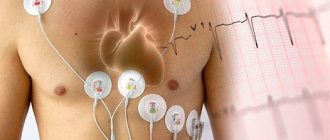
Tachycardia, which is a sign of a disease, is characterized by the following symptoms.
- A feeling of interruptions and/or “failures” in the functioning of the heart is the most common manifestation.
- Shortness of breath (lack of air), which occurs with little physical activity or even at rest.
- Pain in the heart (accompanies an attack of tachycardia in vegetative-vascular dystonia and some other diseases).
- Dizziness and darkening of the eyes, lightheadedness and even fainting (this is due to impaired blood supply to the brain).
In general, the manifestation of clinical symptoms of tachycardia depends on the nature of the underlying disease, as well as on the severity and duration of the arrhythmia itself.
GH, IGF-1 and cardiac contractility
- Most studies indicate that IGF-1 increases myocardial contractility. There is no data on the direct effect of GH (not mediated by IGF-1) on cardiac muscle contractility. There are also paradoxical results: in mutant mice with a reduced concentration of IGF-1, myocardial contractility was increased.
- Changes in intracellular Ca2+ currents
- In in vitro experiments, IGF-1 slows down the release of K+ from the cell, which, in turn, increases the time of Ca2+ influx through L-type Ca2+ channels.
- The results of a number of studies contradicted those described above: a pronounced inotropic effect of IGF-1 was achieved against the background of a reduced peak amount of Ca2+ in the cell and increased sensitivity of contractile elements to Ca2+.
- In animal models, excess GH caused the formation of myosin isoforms with reduced ATPase activity, that is, requiring less energy to perform contraction.
Aspects of the influence of GH on the cardiovascular system
- Risk factors Risks associated with GH deficiency Insulin resistance
- Increased LDL levels
- Slowing down fibrinolysis
- Increased sympathetic activity
- Reducing the volume of extracellular fluid and, as a result, reducing preload on the heart
- Changes in body composition
- Violation of thermoregulation
- An initial decrease in sympathetic activity is observed 1 year after the start of treatment. One study found recovery of insulin sensitivity after 7 years.
- The incidence of cardiovascular mortality is significantly increased in patients with hypopituitarism compared with healthy controls.
Diabetes
Diabetes mellitus leads in mortality from diseases of the circulatory system among other endocrine diseases.
With diabetes mellitus, various lesions of the cardiovascular system develop:
- changes in the blood vessels of the heart - diabetic macroangiopathy;
- arterial hypertension (due to diabetic kidney damage, metabolic syndrome - depending on the type of diabetes);
- diabetic cardiomyopathy with rhythm disturbances and circulatory failure;
- IHD, sometimes with painless angina;
- increased levels of cholesterol and triglycerides in the blood;
- increased risk of ventricular fibrillation, myocardial infarction, and cerebrovascular accidents.
Treatment:
- compensation of carbohydrate metabolism (insulin therapy, hypoglycemic drugs);
- antihypertensive drugs, antiplatelet agents;
- lipid-lowering drugs (statins).
Potential role of GH in the treatment of heart failure
- GH and IGF-1 have been repeatedly shown to improve systolic cardiac function, including in patients with advanced ischemic or dilated cardiomyopathy. In one experiment on mice, an increase in stroke volume was noted even while taking ACE inhibitors
Causes of tachycardia
An increased or increased heart rate does not always mean there are problems with the cardiovascular system. For example, in preschool age it is considered normal and does not require special help. Symptoms of cardiac tachycardia can also appear in practically healthy people as a result of activation of physiological compensatory mechanisms, that is, as the body’s response to the influence of one or another external factor. The reaction of the nervous and cardiovascular systems, accompanied by the release of adrenaline into the blood, causes increased heart rate. The occurrence of tachycardia can be provoked by the following factors:
- stress, physical activity and emotional arousal;
- increase in ambient temperature;
- consumption of caffeinated drinks, alcohol, and certain medications;
- sudden change in body position, etc.
When the action of the provoking factor ceases, the heart rhythm gradually returns to normal.
However, tachycardia often accompanies the presence of certain pathological conditions. It can be a manifestation of various cardiovascular diseases - arterial hypertension, myocardial infarction, heart disease (rheumatic or congenital), cardiosclerosis, etc. In addition, tachycardia can be neurogenic in nature, that is, associated with disturbances in the functioning of the autonomic nervous system and brain. Other causes include fever that develops against the background of an infectious-inflammatory process (pneumonia, tonsillitis, etc.).
Bibliography
References
- Sacca L, Cittadini A, Fazio S. Growth Hormone and the Heart. Endocr Rev. 1994;15(5):555-573. doi:10.1210/edrv-15-5-555.
- Sverrisdóttir YB, Elam M, Herlitz H, Bengtsson B-Å, Johannsson G. Intense Sympathetic Nerve Activity in Adults with Hypopituitarism and Untreated Growth Hormone Deficiency. J Clin Endocrinol Metab. 1998;83(6):1881-1885. doi:10.1210/jcem.83.6.4895.
- Isgaard J, Arcopinto M, Karason K, Cittadini A. GH and the cardiovascular system: an update on a topic at heart. Endocrine. 2014:25-35. doi:10.1007/s12020-014-0327-6.
- Pete G, Hu Y, Walsh M, Sowers J, Dunbar JC. Insulin-like growth factor-I decreases mean blood pressure and selectively increases regional blood flow in normal rats. Proc Soc Exp Biol Med. 1996;213(2):187-192.
- Donath MY, Jenni R, Brunner HP, et al. Cardiovascular and metabolic effects of insulin-like growth factor I at rest and during exercise in humans. J Clin Endocrinol Metab. 1996;81(11):4089-4094. doi:10.1210/jcem.81.11.8923865.
- Tsukahara H, Gordienko DV, Tonshoff B, Gelato MC, Goligorsky MS. Direct demonstration of insulin-like growth factor-I-induced nitric oxide production by endothelial cells. Kidney Int. 1994;45(2):598-604.
- Haylor J, Singh I, el Nahas AM. Nitric oxide synthesis inhibitor prevents vasodilation by insulin-like growth factor I. Kidney Int. 1991;39(2):333-335.
- Standley PR, Zhang F, Zayas RM, et al. IGF-I regulation of Na(+)-K(+)-ATPase in rat arterial smooth muscle. Am J Physiol Endocrinol Metab. 1997;273(1):E113-E121. https://ajpendo.physiology.org/content/273/1/E113. Accessed August 22, 2015.
- Tivesten Å, Barlind A, Caidahl K, et al. Growth hormone-induced blood pressure decrease is associated with increased mRNA levels of the vascular smooth muscle KATP channel. J Endocrinol. 2004;183(1):195-202. doi:10.1677/joe.1.05726.
- Sverrisdottir YB, Elam M, Caidahl K, Soderling AS, Herlitz H, Johannsson G. The effect of growth hormone (GH) replacement therapy on sympathetic nerve hyperactivity in hypopituitary adults: a double-blind, placebo-controlled, crossover, short-term trial followed by long-term open GH replacement in hypopituitary adults. J Hypertens. 2003;21(10):1905-1914. doi:10.1097/01.hjh.0000084757.37215.55.
- Rosen T, Eden S, Larson G, Wilhelmsen L, Bengtsson BA. Cardiovascular risk factors in adult patients with growth hormone deficiency. Acta Endocrinol (Copenh). 1993;129(3):195-200.
- Borson-Chazot F, Serusclat A, Kalfallah Y, et al. Decrease in carotid intima-media thickness after one year growth hormone (GH) treatment in adults with GH deficiency. J Clin Endocrinol Metab. 1999;84(4):1329-1333. doi:10.1210/jcem.84.4.5595.
- Pfeifer M, Verhovec R, Zizek B, Prezelj J, Poredos P, Clayton RN. Growth hormone (GH) treatment reverses early atherosclerotic changes in GH-deficient adults. J Clin Endocrinol Metab. 1999;84(2):453-457. doi:10.1210/jcem.84.2.5456.
- Penney DG, Dunbar JCJ, Baylerian MS. Cardiomegaly and haemodynamics in rats with a transplantable growth hormone-secreting tumor. Cardiovasc Res. 1985;19(5):270-277.
- Mosca S, Paolillo S, Colao A, et al. Cardiovascular involvement in patients affected by acromegaly: an appraisal. Int J Cardiol. 2013;167(5):1712-1718. doi:10.1016/j.ijcard.2012.11.109.
- Otsuki M, Kasayama S, Yamamoto H, et al. Characterization of premature atherosclerosis of carotid arteries in acromegalic patients. Clin Endocrinol (Oxf). 2001;54(6):791-796.
- Ito H, Hiroe M, Hirata Y, et al. Insulin-like growth factor-I induces hypertrophy with enhanced expression of muscle specific genes in cultured rat cardiomyocytes. Circulation. 1993;87(5):1715-1721.
- Bruel A, Oxlund H, Nyengaard JR. Growth hormone increases the total number of myocyte nuclei in the left ventricle of adult rats. Growth Horm IGF Res. 2002;12(2):106-115.
- Li Q, Li B, Wang X, et al. Overexpression of insulin-like growth factor-1 in mice protects from myocyte death after infarction, attenuating ventricular dilation, wall stress, and cardiac hypertrophy. J Clin Invest. 1997;100(8):1991-1999. doi:10.1172/JCI119730.
- Cuneo RC, Salomon F, Wiles CM, Hesp R, Sonksen PH. Growth hormone treatment in growth hormone-deficient adults. II. Effects on exercise performance. J Appl Physiol. 1991;70(2):695-700.
- Sacca L, Napoli R, Cittadini A. Growth hormone, acromegaly, and heart failure: an intricate triangulation. Clin Endocrinol (Oxf). 2003;59(6):660-671. doi:10.1046/j.1365-2265.2003.01780.x.
- Freestone NS, Ribaric S, Mason WT. The effect of insulin-like growth factor-1 on adult rat cardiac contractility. Mol Cell Biochem. 1996;163-164:223-229.
- Stromer H, Cittadini A, Grossman JD, Douglas PS, Morgan JP. Intrinsic cardiac muscle function, calcium handling and beta -adrenergic responsiveness is impaired in rats with growth hormone deficiency. Growth Horm IGF Res. 1999;9(4):262-271. doi:10.1054/ghir.1999.0117.
- Cittadini A, Ishiguro Y, Stromer H, et al. Insulin-like growth factor-1 but not growth hormone augments mammalian myocardial contractility by sensitizing the myofilament to Ca2+ through a wortmannin-sensitive pathway: studies in rat and ferret isolated muscles. Circ Res. 1998;83(1):50-59.
- Mayoux E, Ventura-Clapier R, Timsit J, Behar-Cohen F, Hoffmann C, Mercadier JJ. Mechanical properties of rat cardiac skinned fibers are altered by chronic growth hormone hypersecretion. Circ Res. 1993;72(1):57-64.
- Drake WM, Howell SJ, Monson JP, Shalet SM. Optimizing gh therapy in adults and children. Endocr Rev. 2001;22(4):425-450. doi:10.1210/edrv.22.4.0438.
- Li L, Ren W, Li J, et al. Increase in serum pregnancy-associated plasma protein-A is correlated with increase in cardiovascular risk factors in adult patients with growth hormone deficiency. Endocrine. 2012;42(2):375-381. doi:10.1007/s12020-012-9697-9.
- Gazzaruso C, Gola M, Karamouzis I, Giubbini R, Giustina A. Cardiovascular risk in adult patients with growth hormone (GH) deficiency and following substitution with GH—an update. J Clin Endocrinol Metab. 2014;99(1):18-29. doi:10.1210/jc.2013-2394.
- Svensson J, Bengtsson BA, Rosen T, Oden A, Johannsson G. Malignant disease and cardiovascular morbidity in hypopituitary adults with or growth without hormone replacement therapy. J Clin Endocrinol Metab. 2004;89(7):3306-3312. doi:10.1210/jc.2003-031601.
- Hartman ML, Xu R, Crowe BJ, et al. Prospective safety surveillance of GH-deficient adults: comparison of GH-treated vs untreated patients. J Clin Endocrinol Metab. 2013;98(3):980-988. doi:10.1210/jc.2012-2684.
- Jin H, Yang R, Gillett N, Clark RG, Ko A, Paoni NF. Beneficial effects of growth hormone and insulin-like growth factor-1 in experimental heart failure in rats treated with chronic ACE inhibition. J Cardiovasc Pharmacol. 1995;26(3):420-425.
- Perrot A, Ranke MB, Dietz R, Osterziel KJ. Growth hormone treatment in dilated cardiomyopathy. J Card Surg. 2001;16(2):127-131.
33. Cittadini A, Saldamarco L, Marra AM, et al. Growth hormone deficiency in patients with chronic heart failure and beneficial effects of its correction. J Clin Endocrinol Metab. 2009;94(9):3329-3336. doi:10.1210/jc.2009-0533.