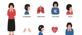
Unpleasant sensations during VSD have different characteristics. A person may experience pressure surges, pain in the heart, rapid heartbeat, anxiety, difficulty breathing, increased body temperature, disturbances in the gastrointestinal tract, restless sleep with VSD, etc. One of the manifestations of VSD is tinnitus. It may also indicate the development of another disease (otitis media or cervical osteochondrosis), and therefore is subject to careful diagnosis. Information about what kind of pain during VSD worries the patient will help the doctor suspect the correct diagnosis, which is confirmed by examination.
Reliable diagnosis and high-quality treatment of VSD in Moscow is performed at the Yusupov Hospital. Extensive experience and high qualifications of doctors allow us to identify the causes of the disease and successfully eliminate it.
Causes of ear congestion
In addition to swelling caused by a cold, ears can become blocked due to some physical reasons:
- Pressure changes when flying on an airplane, riding in an elevator, riding on attractions;
- Getting water into the ear canal while swimming or diving;
- Rhinosinusopathy due to hormonal imbalance during pregnancy.
Also, conditions such as cerumen plug, deformation of the nasal septum, foreign body in the ear, hypertension, taking medications, osteochondrosis, etc. can be the causes of ear congestion.
Insomnia
Insomnia and drowsiness with VSD are common clinical manifestations of nervous system dysfunction. As a result of insomnia, a person becomes irritable, lethargic, and develops chronic fatigue. Facial swelling with VSD can also be the result of insomnia.
The following are the main triggers for insomnia:
- stress;
- neurosis;
- depression;
- change of time zones;
- some medications.
Insomnia with VSD requires urgent treatment, since a prolonged lack of sleep and proper rest causes significant disruption of the functioning of the organs and systems of the body. Insomnia can cause exhaustion of the body - a serious condition that requires complex treatment.
Symptoms of ear congestion
When the ear is blocked, a person’s well-being does not change, but he begins to perceive his own voice differently, external sounds seem muffled, and sudden changes in body position may be accompanied by dizziness and nausea.
If the sound is not perceived in full due to a mechanical obstacle, accumulation of sulfur, narrowing of the ear canal, etc., additional symptoms will be the same as when water gets in.
More complex problems associated with the spine and increased blood pressure can provoke:
- Noise and ringing in the ears;
- Feeling of heaviness in the head;
- Headaches and dizziness.
With Meniere's syndrome and otosclerosis, hearing decreases gradually.
Lower back pain
The cause of back pain during VSD, in particular in the lower back, is most often osteochondrosis of the lumbar region. Degenerative-dystrophic disease of the spinal tissue disrupts the functioning of the nerve roots, which, in turn, begin to transmit incorrect signals to the peripheral nerve endings. Thus, osteochondrosis causes symptoms of VSD.
Back pain with osteochondrosis with the VSD clinic has the following manifestations:
- increased heart rate;
- numbness of the limbs;
- heartburn, nausea;
- lack of air (a cough is often associated with VSD).
Osteochondrosis can develop not only in the lumbar area, but also affect other areas of the spine: the thoracic and cervical regions. VSD and tension in the neck are a consequence of cervical osteochondrosis. Its other symptoms are headache, dizziness, memory impairment, and constant fatigue.
When to go to the ENT doctor?
Constant ear congestion, even without pain, is a cause for concern. Treatment must be directed at the cause, which may be very serious. In severe cases, the patient may require surgery.
It is necessary to consult a doctor in cases where the feeling of congestion does not go away for a long time, the body temperature rises, there is pain and dizziness, any fluids are discharged from the ear, if the hearing loss was preceded by an injury or serious illness.
Treatment of tinnitus with VSD in Moscow
In Moscow, VSD therapy is successfully performed at the Yusupov Hospital. Since VSD is more often a manifestation of some disorder in the physical or psychological state of a person, the pathology requires careful diagnosis. The modern equipment of the Yusupov Hospital allows for examinations of any complexity, which contributes to a quick and accurate diagnosis. Based on the examination data, the doctor (therapist, neurologist or other specialist) creates the most effective therapy that will eliminate unpleasant symptoms and cure the true cause of the pathology.
You can make an appointment with the clinic’s specialists by calling the Yusupov Hospital.
Read also
Adenoids
Probably, in modern times there is not a single parent who has not heard at least once about adenoids.
This is the first question that is asked at an appointment with an ENT doctor: “Do we have adenoids?” What are adenoids?… Read more
Pharyngitis
What is pharyngitis? Pharyngitis is an inflammation of the pharynx, which is located at the back of the throat. Most often it is simply called “sore throat.” Pharyngitis can also cause a sore throat and difficulty...
More details
Hearing loss
Hearing loss is an impairment in the perception of environmental sounds. This disorder occurs in most cases gradually, patients do not immediately understand what is happening and therefore not always in a timely manner...
More details
Nasal congestion in a child
What causes nasal congestion in a child Nasal congestion in a child is a common occurrence. Most often, this problem is caused by viral infections, such as acute respiratory viral infections, influenza, and less commonly, nasal septum defects,…
More details
Runny nose
What is a runny nose? A runny nose is an excessive secretion of mucus from the nasal cavity, caused by its inflammation. The manifestations of a runny nose can range from clear fluid to thick mucus. Source...
More details
Tinnitus is a phantom sound perception in the absence of an objective, external acoustic stimulus [1]. Ear noise significantly worsens the patient’s quality of life, as it is often accompanied by hearing loss, hyperacusis, impaired attention, irritability, insomnia, anxiety, and depression.
Ear noise is heterogeneous, and therefore the diagnosis of this pathology requires an integrated interdisciplinary approach. Differential diagnosis should be based on identifying subgroups with established causes of the disease. First of all, it is necessary to distinguish between groups of subjective and objectively auscultated tinnitus.
Subjective ear noise occurs in 5-15% of the population [2, 3], while objective ear noise is much less common.
Typically, objective noise is characterized as a sound that is heard not only by the patient, but also by others. However, it can be viewed from another point of view: objective - as having some additional source of sound generation. Such a source of sound can be pathological blood flow in the vessels located near the middle ear, or myoclonus of the muscles of the middle ear and topographically close areas.
Constant vascular noise is observed with arteriovenous malformations, as well as with vascular tumors of the middle ear [4, 5]. Transient vascular tinnitus may be caused by medications, hypertension, anemia, or intercurrent illnesses such as migraine.
For the differential diagnosis of pulsating tinnitus and the choice of neuroimaging method, examination by an otorhinolaryngologist plays a vital role. Detection of signs of a neoplasm during otoscopy requires first of all to exclude the presence of a vascular tumor in the middle ear. The most common tumor is a paraganglioma.
Paragangliomas (chemodectomas, glomus tumors) are mainly benign tumors of neuroectodermal origin. The source of chemodectome growth is specialized tissue with regulatory functions, concentrated in the paraganglia. Paraganglia are part of the diffuse neuroendocrine system; they are located in close contact with the nerves and vessels of the head and neck: in the bifurcation of the carotid artery, the bulb of the jugular vein, along the vagus nerve, in the tympanic cavity. The areas of primary growth of paraganglioma are called carotid, jugular, vagal and tympanic. The term tympanojugular paraganglioma is used in cases where a tumor that primarily arose in the jugular vein grows into the tympanic cavity, or when it is impossible to determine the site of primary growth of a large neoplasm.
Paragangliomas are characterized by a high degree of vascularization, infiltrative, locally destructive, and slow growth. Clinical manifestations of tympanic paraganglioma (TP) depend on the size and spread of the tumor. Early symptoms include pulsating tinnitus, aggravated by physical activity, and hearing loss [7]. In some cases, TP actively produces catecholamines [8], which is clinically manifested by uncontrolled hypertension [9].
The further development of the symptoms of the disease depends on the size of the tumor and the direction of its growth. Usually T.P. spreads along the path of least resistance: into the auditory tube, mastoid cells, along preformed pathways, i.e. perivascular and perineural. As it grows, jugular paraganglioma destroys the bottom of the tympanic cavity, involving its contents in the process with subsequent destruction of the medial wall, causing dizziness, sensorineural hearing loss, and facial nerve paralysis.
In the absence of middle ear pathology, it is desirable to determine whether the source of tinnitus is a pathology of the arterial or venous segment of the cerebral circulation. Presumably, the topic of the lesion in this case can be judged by the decrease in noise when the ipsilateral carotid artery or ipsilateral jugular vein is clamped [6]. Depending on the results obtained, a visualization method is chosen: CT/MRI arterio- or venography.
Objective muscular tinnitus is based on involuntary, irregular muscle contractions (myoclonus), which are perceived by the patient as clicks in the ear.
The source of muscle tinnitus is most often tremor (myoclonus) of the soft palate or myoclonus of the middle ear. However, in the literature there are isolated descriptions of ear noise associated with myoclonus of the external muscles of the ear and head muscles - m . temporalis
and
m .
occipitalis [10].
Myoclonus of the soft palate is manifested by rhythmic, uncontrolled contractions. In the diagnosis of myoclonus of the soft palate, it is important to differentiate between symptomatic myoclonus, caused by a lesion in the pons or cerebellum, and essential myoclonus, which occurs in patients without intracranial pathology [11].
Clinical manifestations of tremor of the soft palate are the sensation of involuntary movements of the soft palate in combination with rhinolalia or without it - in 20% of patients, clicks in the ear - in 46.7% of patients, or the presence of both symptoms - in 33.3% of patients. In 53.3% of patients, examination reveals movements of the pharyngeal muscles synchronous with the soft palate [12]. Tremor of the soft palate persists during sleep, but disappears when swallowing and with the patient in the supine position. It is believed that objective ear noise in the form of clicks in the ear with tremor of the soft palate is caused by secondary movements of the walls of the auditory tube [13].
Myoclonus of the middle ear muscles is another possible cause of objective ear noise. This term has been proposed to denote tinnitus resulting from dysfunction of one or both intraauricular muscles: m . tensor tympani
and
m.
stapedius .
This type of pathology can be established by characteristic clinical characteristics (feeling of clicking in the ear), as well as on the basis of impedance measurements and otomicroscopy (with myoclonus m . tensor tympani
).
Myoclonus of the muscles of the auricle is extremely rare. In the literature, this phenomenon is characterized by different terms: ear tic, moving ear syndrome, muscular dystonia, ear dyskinesia [14]. Myoclonus of the muscles of the auricle is often accompanied by its visible rhythmic movement; as a rule, it disappears during sleep and when holding the breath.
Treatment of objective muscular tinnitus is very difficult. One of the areas of treatment is pharmacotherapy, but the assessment of its effectiveness is very controversial, and the evidence base is insufficient [3]. Attempts to treat this group of patients by prescribing anxiolytics, antiepileptic drugs, antidepressants, and using masking white noise turned out to be ineffective. Clinically significant results were obtained when botulinum toxin was injected into the soft palate, causing “chemical denervation” of the muscles for several weeks [15, 16, 17]. Botulinum toxin inhibits the release of acetylcholine from presynaptic nerve terminals, causing "chemical denervation" of the muscle for several weeks. When botulinum toxin is injected into the soft palate, side effects are possible in the form of open nasal sounds, velopharyngeal insufficiency leading to ingested food entering the nasal cavity, dysphagia, as well as subjective increased noise in the opposite ear as a result of the unmasking effect. Usually these adverse events disappear within 10-14 days. The severity of side effects can be minimized by targeted administration of botulinum toxin to the area of maximum myoclonic activity under electromyography control [16, 18]. The dose of the drug should be selected individually for each patient using a titration method [16, 17].
The regimen for administering botulinum toxin depends on the patient’s predominant complaint: for tinnitus, the drug is administered transorally into the m. tensor veli palatini
in the lateral part of the soft palate medial to the projection of the hook of the pterygoid process, if the sensation of involuntary movements of the soft palate predominates, the injection is performed on both sides of
the uvula
[17]. The dose and points of subsequent injections depend on the effect of the first injection and the zone of predominance of muscle contractions.
To relieve ear noise caused by myoclonus of the muscles of the auricle, a good effect is achieved by administering botulinum toxin according to the scheme proposed by K. Lee et al. [19]: 10 units. botulinum toxin to the area m. auricularis posterior
and 10 units.
- to area m.
temporalis [20].
If conservative treatment of myoclonus of the intraauricular muscles is ineffective, surgical intervention is possible - selective tenotomy of the affected muscle.
The causes of subjective ear noise are very diverse (Table 2).
Table 2. Causes of subjective tinnitus
Paroxysmal tinnitus can be a manifestation of Meniere's disease [21], dehiscence syndrome of the superior semicircular canal [22], migraine, epilepsy, vasoneural conflict of the auditory nerve. In these cases, differential diagnosis may require MRI, auditory evoked potentials, vestibular tests, and EEG.
Persistent, non-pulsatile ear noise may be associated with conductive or sensorineural hearing loss. In the presence of conductive hearing loss and low-pitched noise, otosclerosis, various forms of otitis, and dysfunction of the auditory tube should be excluded. Sensorineural hearing loss is characterized by high-pitched noise; in these cases, additional examination of the patient is required for unilateral lesions in order to exclude acoustic neuroma and other tumors of the cerebellopontine angle.
If ear noise is accompanied by headache, then the patient’s examination should be aimed at excluding a space-occupying process, benign intracranial hypertension, cerebrospinal fluid circulation disorders, and craniocervical anomalies. In cases of paroxysmal headache of the hemicrania type, combined with the appearance of homolateral noise in the ear, one should remember the presence of Slader's syndrome [23], since the pterygopalatine ganglion plays a leading role in the ventilation function of the auditory tube. With ganglioneuritis of the pterygopalatine ganglion, the function of the auditory tube is observed, which is manifested by a feeling of ear fullness and the appearance of low-pitched tinnitus.
When collecting complaints and anamnesis, it is necessary to note the following characteristics of ear noise: duration of persistence of the symptom; initial appearance of noise (gradual or acute); what the patient associates with the appearance of noise (acoustic, traumatic brain or whiplash injury, hearing loss, dizziness, hearing loss); nature of the noise: constant or intermittent, paroxysmal, pulsating (synchronously with the heartbeat), fluctuating; unilateral, bilateral (symmetrical or asymmetrical), noise inside the head; noise characteristics: low- or high-pitched.
Depending on the answers to these questions, 2 groups of patients can be distinguished. The first group is patients with persistent, long-lasting ear noise, who require a general clinical examination, hearing testing, and treatment as planned.
The second group of patients who exhibit the presence of “red flags” [2] require immediate examination and treatment. These are patients with acute pulsating noise, including after traumatic brain injury; with noise that occurred simultaneously with acute hearing loss; with unbearable noise, accompanied by depression.
A pulsating noise that occurs after a traumatic brain injury may indicate the formation of a carotid-cavernous anastomosis (CCJ). The CCS is a pathological communication between the internal carotid artery or one of its branches and the cavernous sinus, through which arterial blood is discharged into the venous system.
Clinical symptoms of CCS are caused by hemodynamic disturbances. Arterial blood through the formed anastomosis rushes against the blood flow into the cavernous sinus and the veins flowing into it, causing increasing insufficiency of blood supply to the brain and venous stagnation in the orbit. In this case, the cavernous sinus stretches, compressing the cranial nerves passing through it. CCS is manifested by a typical triad of symptoms: proptosis, injection of conjunctival vessels and noise in the head, synchronous with the pulse and decreasing when the ipsilateral carotid artery is clamped [24, 25]. This triad can be supplemented by headache, exophthalmos with visible or palpable pulsation of the eyeball, chemosis, diplopia, ophthalmoplegia, increased intraocular pressure and decreased vision [26]. On auscultation, a pulsating noise is heard in the orbital area, synchronous with the pulse. Occasionally, the carotid-cavernous anastomosis is not accompanied by pulsating exophthalmos, and sometimes there is no vascular murmur.
Natural variations in the structure of the venous system of the skull affect the clinical manifestations of CCS: in patients with a wide superior or inferior petrosal sinus, arterial blood may be discharged into the internal jugular vein, which is manifested by a decrease in the severity or absence of ocular symptoms ( eng.:
"white eye shunt") With a decrease in the functionality of the stony sinuses, blood discharge occurs into the veins of the cerebral cortex, nasal cavity and eyeball, which creates the risk of developing cerebral edema, cerebrovascular hemorrhages and profuse, life-threatening nosebleeds [5, 27].
In the diagnosis of CCS, modern imaging methods are of great importance, revealing the expansion of the cavernous sinus, congestion in the veins of the orbit, pia mater and cerebral cortex. The gold standard for diagnosing CCS is digital subtraction angiography [28]. MRI can detect swelling of the orbital and cerebral tissues, as well as abnormal blood flow in the cavernous sinus [29], and Doppler ultrasound can detect pulsatile or reverse blood flow in the superior orbital artery.
Acute unilateral hearing loss of the sensorineural type is accompanied by the appearance of tinnitus, and in some cases, dizziness. As a rule, this is a manifestation of labyrinthine infarction. The prospect of hearing restoration in this group of patients depends on how quickly treatment is started.
Patients with unbearable ear noise combined with depression also require immediate treatment measures due to the high risk of suicide attempts.
Subjective tinnitus is usually combined with hearing loss (conductive, perceptive, or mixed). Depending on the form of hearing loss and the degree of its severity, a large number of methods for correcting impaired auditory perception and accompanying ear noise have been proposed: pharmacotherapy, acupuncture, hirudotherapy, psychotherapy, physiotherapy, sound and music therapy, hearing aids (traditional, partially implantable hearing aids and cochlear implantation), non-invasive neuromodulation of brain stem structures and others [30], however, all of them can, at best, reduce the intensity of tinnitus, but not relieve the patient from this painful symptom.
The results of modern studies show that with tinnitus, regardless of its original cause, the auditory system is involved in processes that occur in its central parts, and is not accompanied only by morphological changes in the cochlea, as previously thought [31]. The main cause of tinnitus is considered to be increased spontaneous activity of neurons in the auditory cortex of the brain and pathological hypersynchronization of neuronal activity in the auditory system, or a combination of both factors [32].
Based on theoretical concepts according to which subjective tinnitus is a consequence of morphofunctional disorders in the auditory system, research on the treatment of tinnitus in the direction of optimizing the functioning of neural networks and reducing sensory hyperactivity has received new impetus, i.e., taking into account the effects of the implementation of neuroplasticity mechanisms [33] . Such properties are possessed by drugs that have a metabolic and neuroprotective effect on the central nervous system. One such drug is EGb 761 (memoplant).
EGb 761 (memoplant) is a standardized extract of Ginkgo biloba leaves with a strictly defined, stable and precise chemical composition. The main pharmacological properties of this drug are an increase in tissue blood flow (both cerebral and peripheral), a decrease in blood viscosity, replenishment of the deficiency of certain neurotransmitters and growth factors, and elimination of free radicals [34].
The literature contains a large number of clinical studies, including randomized, double-blind, placebo-controlled, which confirmed the effectiveness of EGb 761 in a wide range of diseases of the nervous system: mild cognitive impairment [35, 36], asthenic symptom complex [37], treatment of chronic vascular diseases of the brain [38] and vertebrobasilar insufficiency [39].
The influence of EGb 761 on metabolic processes in nervous tissue was the basis for conducting experimental and clinical studies of the possibility of its use for the treatment and prevention of disorders of the auditory analyzer. It has been established that the neuroprotective effect of EGb 761 may be due to the regulation of phosphorylation of the pAkt protein [40], which affects the adequate expression of proteins-inducers and inhibitors of apoptosis, which ensures greater cell survival in response to damage. In addition, the introduction of EGb 761 limits the release of caspase-3, which is one of the key factors in the induction of apoptosis. The neuroprotective effect of EGb 761 ensures increased resistance of nervous tissue to a variety of damaging factors. Thus, experiments on animals showed a decrease in the ototoxic effect of cisplatin [41] and a decrease in the damaging effect of noise on the auditory analyzer [42, 43] while taking the drug. These effects reflect neuroplasticity processes and help explain the additive therapeutic effect of EGb 761 in patients with sudden sensorineural hearing loss treated with corticosteroids, as was shown in a randomized controlled trial by J. Koo et al. [44]. A similar result was reported in the treatment of blast trauma victims [45].
The obtained data on the effect of EGb 761 on the structural and functional state of the nervous system in the form of stimulation of neuroplasticity processes served as a theoretical basis for studying the possibility of its use for the treatment of chronic subjective tinnitus. In a randomized, controlled, double-blind study, C. Morgenstern and E. Biermann [46] showed the effectiveness of using the drug EGb 761 at a dose of 80 mg 2 times a day. The first significant differences in the level of ear noise between the experimental and control groups were noted after 8 weeks of treatment and reached a maximum 12 weeks after the start of taking the drug. Thus, this study demonstrated a dose-dependent effect of EGb 761.
The problem of the dose-dependent effect of EGb 761 was addressed in a meta-analysis presented in the Cochrane Database, which included studies published before 2006 [47]. It turned out that patients receiving the drug for at least 24 weeks (at least 240 mg per day) had a more significant positive effect compared to patients receiving placebo, while lower doses had a significantly less effect, which confirmed assumption of a dose-dependent effect of the drug. It was noted that improvement in cognitive functions was recorded already at the 12th week of treatment, and when treatment was continued until the 24th week, there was no additional increase in effectiveness. These data were confirmed in a 2015 Cochrane review [48].
Of no less importance is the effect of EGb 761 on emotional and behavioral disorders: a significant decrease in such manifestations as apathy, irritability, dysphoria, dissomnia, depression was noted when taking the drug at a dose of 240 mg per day [49-51], which is extremely important for patients suffering from ear infections. noise. The effect of EGb 761 on emotional and behavioral disorders may be due to its effects on serotonin and glucocorticoid receptors in the hippocampus [52].
Thus, ear noise is a symptom that can signal the presence of serious diseases that threaten the patient’s life. Such patients require careful examination and treatment that can improve their quality of life.
The author declares no conflict of interest.