Pathological conditions that are manifested by changes in the frequency and strength of heart contractions. They manifest themselves as pain in the heart, rapid heartbeat, irregular heartbeat, shortness of breath and dizziness. Heart conduction disturbances (blocks) are a common finding on electrocardiographic (ECG) examinations. Most often, they do not manifest themselves clinically, but some blockades require the implantation (installation) of a permanent pacemaker (pacemaker).
Many types of intracardiac blockades (for example, incomplete blockade of the right bundle branch) are a normal variant.
Cardiological examination for cardiac conduction disorders is intended not only to determine the type of blockade, but also to determine whether it is a manifestation of organic heart damage. In addition, not all cases of blockade need to be treated. The main indications for installing a pacemaker are fainting and pre-fainting conditions, but you need to be sure that fainting is caused precisely by cardiac conduction disorders.
General picture of the disease
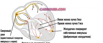
The structure of the His bundle involves the presence of two anterior legs and a posterior one. The branch on the right has a wide bundle that penetrates the muscle layers of the right ventricle. To understand what right heart branch block is, it is necessary to consider the features of the changes observed in this case:
- Throughout the cardiac system, the conduction of impulses slows down.
- The right parts of the organ are excited, affecting the septum present between the ventricles.
- The unblocked left ventricle is involved in the process, after which the right one is also excited.
Impaired conduction along the right bundle branch can negatively affect the functioning of the ventricles, as a result of which the time of their excitation changes. As a result, loss of normal performance of the right departments is possible.
Against the background of the development of this pathology, impulses can reach the left bundle of His without changes, without spreading to the right leg. Due to this disconnection in the functioning of the parts of the heart, the development of serious pathologies of the organ is possible.
Diagnosis of cardiac conduction disorders (part 3)
The ECG shows signs of RBBB (QRS complex more than 0.1 s, with a predominant R wave in leads V1–V2, III, aVF) in combination with signs of LBBB (QRS complex more than 0.1 s, with a predominant R wave in leads V5–V6 , I, aVL), they are presented in Figure 27. 4.11. Three-fascicular block is a conduction disorder along the right leg in combination with intermittent blockade of the anterior and posterior branches of the left leg. ECG signs: 1. Block of the right bundle branch in combination with blockade of the anterior and posterior branches of the left bundle branch (RBBB + RBBB/BLBB). 2. Block of the right bundle branch in combination with blockade of the anterior branch of the left bundle branch and AV block of the I–II degree (RBBB + RBBB + AV block of the I–II degree). 3. Block of the right bundle branch in combination with blockade of the posterior branch of the left bundle branch and AV block of the I–II degree (RBBB + BLBB + AV block of the I–II degree) (Fig. 28). 4.12. Arborization blockade is a disorder of conduction along Purkinje fibers. The QRS complex is low-amplitude, widened (more than 0.12 s), jagged (Fig. 29). 4.13. Transient blockade of the branches of the His bundle and the branches of the left leg (transient blockade of the legs and branches of the left leg) is a reversible conduction disorder resulting from organic damage to the myocardium or functional conduction disorders. With transient blockades of the branches of the His bundle, during the disappearance of the blockade, a negative T wave and/or depression of the ST segment are recorded in leads where the QRS complex was negative during intraventricular blockade - “post-blockade syndrome”, a variant of “post-depolarization syndrome” (Fig. 30–32) . 4.14. Exit block is a local block that prevents the excitation impulse (sinus, ectopic, or artificially caused by the pacemaker) from spreading into the surrounding myocardium, despite the fact that the latter is in the extra-refractory period. Output blockade is the result of inhibited conduction near the source of impulse formation or reduced intensity of the excitation impulse. The first mechanism is much more common than the second. Output blockade as a result of myocardial conduction disturbance near the source of impulse formation can be type I with Samoilov-Wenckebach periodicity or type II - sudden onset, without gradual deepening of conduction disturbance. Output blockade is a common phenomenon; it occurs with different localizations of the automatic center (Fig. 33). Conclusion The variety of cardiac conduction disorders makes their diagnosis significantly difficult. However, the relevance of adequate assessment of dysfunction of the cardiac conduction system is beyond doubt. This publication is intended to help primarily the practicing cardiologist, as well as other specialists.
* Part 1, see RMJ. 2013, no. 4. pp. 237–240. Part 2 see RMJ. 2013, no. 12, pp. 647–650.
Literature 1. Cardiac arrhythmias / Ed. W. J. Mandela. M.: Medicine, 1996. P. 512. 2. Janashia P.Kh., Shevchenko N.M., Shlyk S.V. Heart rhythm disturbances. M.: Overley Publishing House, 2006. P. 320. 3. Isakov I.I., Kushakovsky M.S., Zhuravleva N.B. Clinical electrocardiography. L.: Medicine, 1984. 4. Cardiology in questions and answers / Ed. Yu.R. Kovaleva. St. Petersburg: Foliant Publishing House LLC, 2002. P. 456. 5. Kushakovsky M.S. Cardiac arrhythmias. St. Petersburg: Hippocrates, 1992. 6. Kushakovsky M.S., Zhuravleva N.B. Arrhythmias and heart block (atlas of electrocardiograms). L.: Medicine, 1981. 7. Murashko V.V., Strutynsky A.V. Electrocardiography: Textbook. allowance. 3rd ed., revised. and additional M.: Medpress LLC; Elista: APP "Dzhangar", 1998. P. 313. 8. Orlov V.N. Guide to electrocardiography. M.: Medical Information Agency LLC, 1999. 528 p. 9. Tomov L., Tomov I. Heart rhythm disturbances. Sofia: Medicine and Physical Education, 1976. 10. Zimmerman F. Clinical electrocardiography. M.: Binom, 1997. 11. Shchikota A.M., Snetkova A.A., Shekhyan G.G., Yalymov A.A. Clinical problem on the topic: differential diagnosis of diseases accompanied by ST segment elevation, No. 3 (Brugada syndrome ) // Directory of a polyclinic doctor. 2012. No. 5. P. 14–15, 38. 12. Yakovlev V.B., Makarenko A.S., Kapitonov K.I. Diagnosis and treatment of heart rhythm disorders. M.: Binom. Knowledge Laboratory, 2003. P.168. 13. Yalymov A.A., Shekhyan G.G., Shchikota A.M. Guide to electrocardiography / Ed. Zadionchenko V.S. Saarbrucken, Germany. Publisher: LAP LAMBERT Academic Publishing GmbH&Co. KG, 2011. 14. Brugada P., Brugada J. Right bundle branch block, persistent ST segment elevation and sudden cardiac death: a distinct clinical and electrocardiographic syndrome. A multicenter report // J. Am. Coll. Cardiol. 1992. Vol. 20. P. 1391–1396. 15. Rosenbaum MB, Elizari MV, Lazzari JO The Hemiblocks: New Concepts of intraventricular Conduction Based on Human Anatomical, Physiological and Clinical Studies. Florida, 1970.
Causes
Such changes most often occur due to structural changes in the heart muscle. This may be caused by one of the following diseases:
- various types of heart defects;
- ischemia;
- hemochromatosis;
- heart attack, cardiosclerosis and other heart ailments;
- pulmonary heart;
- thromboemolia of the pulmonary arteries;
- amyloidosis;
- poisoning with medications or exceeding recommended doses;
- hypertension.
In some cases, the cause of conduction disturbance may be one of the extracardiac factors:
- systemic connective tissue diseases;
- problems of water and electrolyte nature;
- congenital pathologies associated with the conductive function of the heart.
We should not forget about the characteristics of the body. According to statistics, every twentieth patient has a congenital conduction disorder in the right leg, which is the norm.
Symptoms
Complications in diagnosing the disease arise because the conduction disorder of the right bundle branch does not have any significant manifestations. The patient simply cannot detect this deviation on his own.
Most often, the disease is detected randomly during a routine ECG. However, the patient may have complaints such as pain in the heart, shortness of breath, interruptions in heart rhythm, and severe fatigue, which appear due to the progression of the underlying disease that caused the blockade.
If other sectors are affected, the following symptoms are possible:
- Signs of hemiblockade of the posterior or anterior left branch depend on the underlying disease. They are usually mild and involve heart pain, shortness of breath, and fatigue.
- Complete blockade of the left leg of the heart is manifested by dizziness, heart pain, and palpitations. The appearance of these symptoms can be caused by extensive changes in the left ventricle, including acute infarction.
- Three-fascicle blockade can be characterized by a complete absence of impulse conduction. Patients experience frequent dizziness, interruptions in heart function, and fainting. If adequate treatment is not carried out, various complications are possible, including a heart attack.
Detection of conduction disorders using Holter monitoring.
Akselrod A.S., Head of the Department of Functional Diagnostics,
Cardiology Clinic of the MMA named after.
THEM. Sechenov Conduction disturbances are less common in cardiologist practice than cardiac arrhythmias. However, a significant proportion of syncope of unknown origin is represented by conduction disorders. If they are transient (which happens quite often), it is extremely difficult to detect them when recording a standard ECG. In such a situation, consistent use of a 24-hour recorder over 3 days or a single use of a 72-hour recorder is absolutely recommended.
As is known, patients with various conduction disorders may not present any complaints for a long time. In such situations, the appearance of syncope is often the first indication for Holter ECG monitoring.
During 24-hour ECG recording, it is possible to detect conduction disturbances that occur only at night. Of course, daily ECG monitoring also reveals the connection between conduction disorders and medication use, physical activity, etc. Transient sinoatrial and atrioventricular blockades, transient frequency-dependent blockades of intraventricular conduction, changes in the degree of previously diagnosed blockade - this is an incomplete list of the most common conduction disorders, which can only be identified with long-term ECG monitoring.
When purchasing software, you should pay attention to the mandatory presence of three features:
1. changing the tape drive speed: this feature allows you to more clearly set the boundaries of the PQ interval and PP distance;
2. changing the overall voltage: this feature allows you to increase the amplitude of the P wave and, thus, visualize it more clearly in doubtful cases;
3. the presence of a ruler with colored stretchable jaws: when you set these jaws to the interval you need, its duration in ms automatically appears on the fragment.
Sinoatrial blocks
associated with a slowdown (grade 1) or disruption (grades 2 and 3) of the generation or conduction of sinus node impulses to the atrial myocardium and, accordingly, the atrioventricular node. Sinoatrial block can be transient or permanent, occur at any heart rate and be combined with other conduction and heart rhythm disorders.
Sinoatrial block 1st degree
can be suspected by fragments of a sudden slowdown in rhythm followed by its acceleration (difficult to differentiate with sinus arrhythmia) during Holter monitoring.
With 2nd degree SA blockade
Some of the impulses arising in the sinus node do not reach the atria. In this case, a pause (more than 2 seconds) without atrial activity is recorded on the ECG: unlike AV block, during a pause with SA block there are no P waves.
With 2nd degree block of type I (partial sinoauricular block with Samoilov-Wenckebach periods)
There is a progressive shortening of the RR intervals before a long pause - Samoilov-Wenckebach periodics. In this case, the degree of conduction disturbance can be characterized by the ratio of the number of sinus impulses, for example, 3:2, etc. (in the numerator the number of sinus signals is set
impulses, including the expected and failed impulses; the denominator is the number of actually conducted impulses). The detected pause is not a multiple of the distance PP of the main rhythm.
For sinoatrial block of the 2nd degree, type II (Mobitz type)
no such periodicals are found. This type of blockade is diagnosed more often. The identified pause is a multiple of or equal to one distance PP of the main rhythm. Often, with this type of blockade with a 2:1 or greater degree of blockade, it becomes necessary to differentiate monitoring fragments from sinus bradycardia. Often, during the same Holter registration, it is possible to register both types of SA blockade.
Pay attention to the ability of your software to display in each of the printed fragments both the duration of the pause and the heart rate value against the background of this pause. This marking makes the fragment very visual and once again emphasizes its diagnostic significance (Fig. 1).
Rice.
1. Patient S., 64 years old, variants of sinoatrial block of the second degree: A - C A block of the 2nd degree, type I with Samoilov-Wenckebach periodicity; B – SA blockade 2nd degree type II with 3:2.
A
B
About the third degree of sinoatrial block (complete sinoatrial block or sinus node failure, “sinus arrest”)
they speak in the absence of atrial waves and the presence of replacement contractions from the distal centers of automaticity - the AV connection or the ventricular conduction system (Fig. 2).
Often during Holter monitoring one can see fragments of conduction disturbances that occur against the background of respiratory arrhythmia. In such a situation, it can be quite difficult to qualify the identified pauses. For example, in patient Zh., 45 years old, at night (from 2:00 to 5:00) episodes of SA conduction disturbance were recorded without multiplicity and clear Samoilov-Wenckebach periodicity, 9 pauses of more than 4 seconds, including 2 episode of sinus node arrest.
Fig.2. Patient Zh., 45 years old: A - episodes of slowing of SA conduction without a clear multiplicity and Samoilov-Wenckebach periodicity, B - arrest of the sinus node with the formation of a pause of 4.048 sec.
A
B
For novice doctors, I would like to note three important points:
1. often the degree and type of blockade may vary depending on the time of day;
2. the lack of multiplicity of the PP interval and the duration of pauses may be due to concomitant sinus arrhythmia, often respiratory;
3. when qualifying a pause as an SA blockade, you must be absolutely sure that this fragment is not artificial: the pause is duplicated in both leads. In doubtful cases, monitoring will have to be repeated.
Atrioventricular blockade.
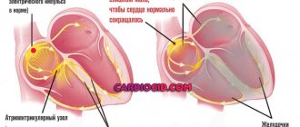
Atrioventricular (AV) blocks are caused by damage to the conduction system at the 2nd and 3rd levels - the conduction of the sinus impulse to the atrioventricular node, as well as pathology of the atrioventricular node itself. In this case, both a delay in the conduction of the impulse from the atria through the AV node, and a complete cessation of its conduction are possible.
Prolongation of the PQ interval by more than 200 ms in adults and more than 170 ms in children indicates 1 degree of AV block (slowing of AV conduction)
. Incidental detection of this type of blockade at night in patients taking beta-blockers and not showing any complaints is one of the most common favorable conduction disorders in practical cardiology and can be qualified in conclusion as “slowing of AV conduction” if PQ does not exceed 300 msec (Fig. 3).
Rice. 3. Patient R., 57 years old: slowing of AV conduction was detected during night sleep (PQ interval reached 240 ms). A – PQ 146 ms (15:10), B – PQ 240 ms (4:33).
A
B
A much greater danger is posed by a significant (more than 300 ms) slowdown in AV conduction, which must be qualified in the conclusion as “1st degree AV block” (Fig. 4). When a PQ interval of more than 300 ms is recorded on the resting ECG, the patient is advised to have 24-hour ECG monitoring to decide on the need for therapy correction. Such severe conduction disturbances often progress over the course of a day.
Fig.4. Patient G, 64 years old: 1st degree AV block
“Loss” of the ventricular complex (pause, a multiple of the duration of the RR interval) with registration of an unchanged P wave (as opposed to sinoatrial block) is a sign of 2nd degree AV block
.
With an increasing lengthening of the PQ interval before the pause, they speak of type I partial AV block of the 2nd degree with Samoilov Wenckebach periods (Mobitz type I)
.
In the absence of such periodicals, type II AV block of the 2nd degree (Mobitz type II)
. It is convenient to indicate the degree of conduction using the ratio 5:2, 3:2, etc. (the first digit indicates the number of P waves, the second - the number of ventricular QRS complexes). It can be extremely useful to use graphs (or tables) of the distribution of pauses by hour. At the same time, having distribution graphs in your program is much more convenient: they are more visual and allow you to quickly and correctly assess the prevalence of pauses by hour (Fig. 5).
Fig.5
.
Patient B, 76 years old: 2nd degree AV block, type II.
A – stereotypical fragment of blockade with the formation of a pause of 2.288 sec; B – graph of the distribution of pauses by hour (predominance at night is expressed) A
B
Complete atrioventricular block (3rd degree AV block, complete transverse block)
is detected as a loss of connection between atrial (P wave) and ventricular contractions (QRS complex), while the atrial rhythm is more frequent than the ventricular one (Fig. 6). In such fragments one can see the layering of P waves on the ventricular QRS complexes, so the possibility of increasing the total voltage (and, accordingly, the amplitude of the P wave) is simply necessary.
Fig.6. 3rd degree AV block in patient Zh., 69 years old.
Often, against the background of 3rd degree AV block, replacement contractions or rhythms are recorded (Fig. 7).
Fig.7.
Patient G, 64 years old: replacement idioventricular rhythm against the background of 3rd degree AV block.
Quite often in patients, AV block occurs sporadically or its degree varies depending on the time of day. Rare episodes of 2nd degree AV block may also occur at night (usually in the early morning hours) with a normal PQ interval for the rest of the monitoring time. In addition, during follow-up of a patient with AV block, one can often see a progressive deterioration of AV conduction over several years (Fig. 8).
Fig.8.
Progressive deterioration of AV conduction in patient L., 45 years old: A – slowing of AV conduction was first detected at the age of 45 years; B – 2nd degree AV block type II at 46 years old; C and D – 2 consecutive episodes of 3rd degree AV block 3:2 and 5:2 with the formation of pauses of 2.31 and 5.34 seconds, respectively.
A
B
IN
G
Every novice doctor is faced with the difficulty of differential diagnosis between 2nd degree AV block type II and 3rd degree AV block. Only with a detailed comparison of the fragments and the use of the “ECG review” option can we conclude that there is a complete transverse blockade on the controversial fragment.
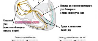
Blockades of the branches of the His bundle
A standard 12-channel resting ECG allows you to clearly diagnose variants of conduction disorders along the His system. During daily ECG monitoring, it is possible to identify transient blockades of the branches of the His bundle, which are recorded at night or, conversely, during intense physical activity. They are often an incidental diagnostic finding. However, such intraventricular conduction disturbances (for example, transient complete block of the left bundle branch) can mimic paroxysmal ventricular arrhythmias and lead to overdiagnosis of life-threatening arrhythmias (Fig. 9).
Fig.9.
Patient K., 72 years old: transient complete blockade of the left bundle branch. A – beginning of the blockade, B – end of the blockade.
A
B
As a rule, it is not difficult to differentiate conduction aberration along the His system from paroxysmal ventricular rhythm disturbances: the block is characterized by a regular regular rhythm, smooth regular cycles, the absence of a compensatory pause (or lengthening of the RR interval) at the end of a rhythm fragment from extended complexes and a smooth restoration of normal sinus rhythm .
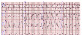
None of the listed signs can be seen in Fig. 10, which allows us to qualify this fragment as ventricular tachycardia. Rice. 10. Patient K., 79 years old: paroxysm of unsustained ventricular tachycardia
In conclusion, I would like to note: for a clear diagnosis of conduction disorders, a single Holter recording is often not enough. If there are dubious changes suspicious for conduction disorders (especially at night), the study must be repeated with a total monitoring duration of up to 72 hours.
Moscow, 04/16/2009
Diagnostic features
Any conduction abnormality can be detected during electrocardiography. Based on the results of this procedure, the specialist determines further actions:
- If incomplete right blockade was detected in the absence of other cardiological diseases, then this can be attributed to the characteristics of the body. Additional research in this case is not prescribed.
- A detected two-fascicle blockade will require in-depth diagnostics. If this deviation has not previously been observed in the patient, then he needs urgent hospitalization, even if there are no complaints. If a complete left blockade exists for a long time, then hospital treatment is not required.
- If a three-fascicular block is detected in a patient, urgent hospitalization is necessary. Here, a full examination will be carried out in a short time to determine the further course of treatment.
Treatment methods
This pathology needs to be eliminated only if the patient has a disease that provoked the development of the blockade. In its absence, therapy is not carried out.
For patients suffering from single- or double-fascicular nonconduction, drug therapy is chosen, which involves taking the following drugs:
- antioxidants;
- vitamins;
- sedative herbal preparations;
- means for eliminating arterial hypertension;
- antibiotics;
- cardiac glycosides and diuretics.
The course of treatment is selected depending on the underlying disease and the stage of its development. In advanced cases, the blockage can be treated surgically. It involves installing a pacemaker for the patient.
The patient’s lifestyle and disease prognosis
If the patient does not have any heart disease, and the blockade of the right leg of the heart occurs in his body without complications, then he can lead a normal life with moderate physical activity. If the pathology is caused by another illness, the patient should limit stressful situations and loads, eliminate bad habits, and monitor nutrition.
If a pacemaker was installed during the operation, the patient should observe the following precautions:
- have with you the identification card of the owner of the pacemaker;
- protect the implantation area from the influence of a mobile phone or electrical appliances;
- undergo an ECG once a year (or more often if there is a special doctor’s order).
Since His bundle block is not an independent disease, but is a consequence of other ailments, the prognosis directly depends on the disease that provoked this pathology. Single-fascicle right blockade without significant heart damage does not pose a danger to human health.
If, as a result of a heart attack, the impulse of the left branch was blocked, then the prognosis is less favorable (mortality is up to 50% with exacerbation of the disease). Three-bundle pathology also has serious consequences, since against its background the likelihood of asystole increases.
The blockade of the His bundle itself is a feature of the body and cannot affect the patient’s quality of life. But with the development of concomitant diseases, the consequences of this pathology can be very dire. To avoid trouble, it is necessary to undergo systematic ECG examinations.
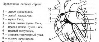
Conduction system of the heart
In general terms, the conduction system of the heart (the system responsible for conducting electrical impulses in the heart) is arranged as follows. The impulses are generated by the sinus node located in the right atrium. Along the intraatrial pathways, these impulses reach the atrioventricular (AV) node, where some delay of the impulses occurs: the atria and ventricles must contract at the same time. The impulse then travels along the bundle branches to the cells (cardiomyocytes) of the ventricles. The bundle of His consists of two legs - right and left. The left bundle branch consists of two branches - anterior and posterior.